. 2014 May 15;9(5):e97826.
doi: 10.1371/journal.pone.0097826. eCollection 2014.

A new restriction endonuclease-based method for highly-specific detection of DNA targets from methicillin-resistant Staphylococcus aureus

Affiliations

A new restriction endonuclease-based method for highly-specific detection of DNA targets from methicillin-resistant Staphylococcus aureus

Maria W Smith et al. PLoS One. .

Abstract

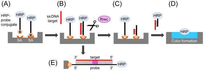
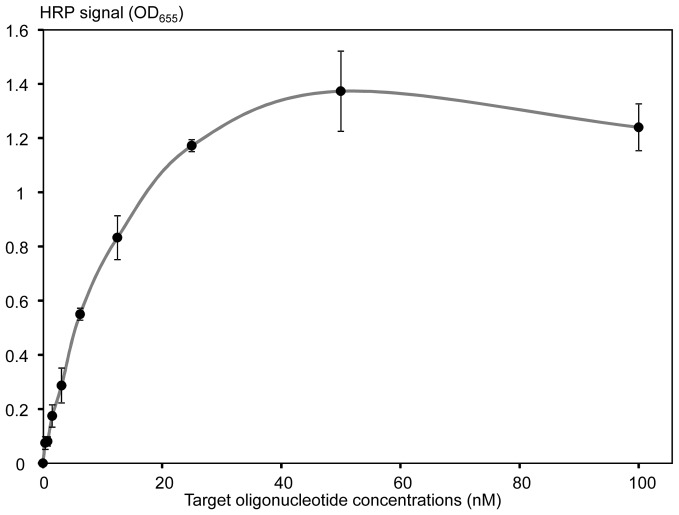
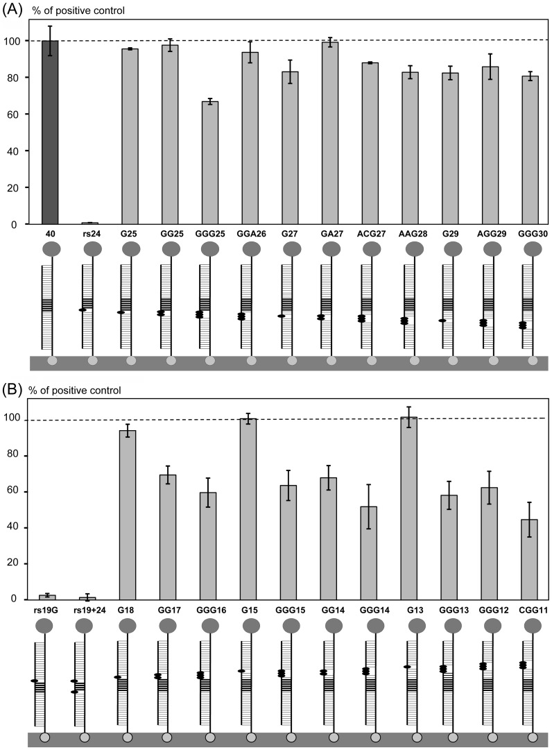
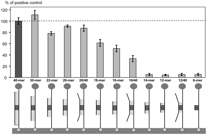
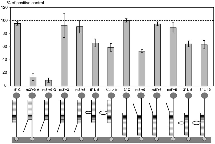
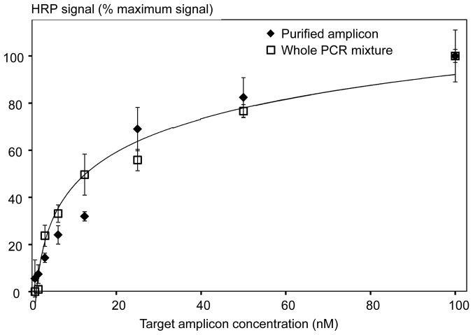
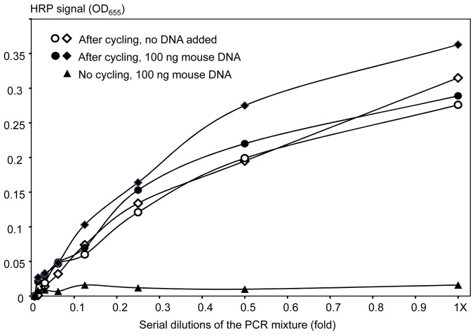
PCR multiplexing has proven to be challenging, and thus has provided limited means for pathogen genotyping. We developed a new approach for analysis of PCR amplicons based on restriction endonuclease digestion. The first stage of the restriction enzyme assay is hybridization of a target DNA to immobilized complementary oligonucleotide probes that carry a molecular marker, horseradish peroxidase (HRP). At the second stage, a target-specific restriction enzyme is added, cleaving the target-probe duplex at the corresponding restriction site and releasing the HRP marker into solution, where it is quantified colorimetrically. The assay was tested for detection of the methicillin-resistant Staphylococcus aureus (MRSA) pathogen, using the mecA gene as a target. Calibration curves indicated that the limit of detection for both target oligonucleotide and PCR amplicon was approximately 1 nM. Sequences of target oligonucleotides were altered to demonstrate that (i) any mutation of the restriction site reduced the signal to zero; (ii) double and triple point mutations of sequences flanking the restriction site reduced restriction to 50-80% of the positive control; and (iii) a minimum of a 16-bp target-probe dsDNA hybrid was required for significant cleavage. Further experiments showed that the assay could detect the mecA amplicon from an unpurified PCR mixture with detection limits similar to those with standard fluorescence-based qPCR. Furthermore, addition of a large excess of heterologous genomic DNA did not affect amplicon detection. Specificity of the assay is very high because it involves two biorecognition steps. The proposed assay is low-cost and can be completed in less than 1 hour. Thus, we have demonstrated an efficient new approach for pathogen detection and amplicon genotyping in conjunction with various end-point and qPCR applications. The restriction enzyme assay may also be used for parallel analysis of multiple different amplicons from the same unpurified mixture in broad-range PCR applications.


Conflict of interest statement

Competing Interests: MS and KS are co-founders of Cascade Biosystems, Inc. AG is an employee of this company. There are no patents, products in development or marketed products to declare. This does not alter the authors' adherence to all the PLOS ONE policies on sharing data and materials, as detailed online in the guide for authors.

Figures

Figure 1
Figure 1. General schematic of the restriction enzyme assay.
(A) Surface immobilization of HRP conjugated to an oligonucleotide probe specific for a target gene of interest. (B) The target DNA (an oligonucleotide or a denatured PCR amplicon) is hybridized to the immobilized probe. (C) Addition of a restriction enzyme (Rrec) that recognizes and cleaves the target-probe ds DNA hybrid, resulting in release of the HRP marker into the reaction solution. (D) The reaction solution is transferred into a new well and mixed with an HRP substrate for colorimetric detection. For each target DNA molecule one HRP molecule is released, resulting in a linear dependence of the signal on the target DNA concentration. (E) Detailed schematic of the double stranded target-probe DNA duplex, with the specific restriction site shown in purple. HRP, horseradish peroxidase; B, biotin; SA, streptavidin.
Figure 2
Figure 2. A typical calibration curve of the restriction enzyme assay generated with a 40-mer oligonucleotide target AMC-40-mer (fully complementary to the MCA-BG probe).
X-axis shows concentrations (nM) of the target oligonucleotide. Y-axis shows the restriction enzyme generated HRP signal that was quantified by the blue color formation as measured by the OD655. The signal values were background-corrected by subtracting the signal generated by the negative control with no target oligonucleotide added. The experiments were performed in triplicate to generate mean values (black circles) and standard deviations (shown with error bars).
Figure 3
Figure 3. Effect of point mutations introduced into the target sequence.
(A) Single, double and triple mutations were introduced between the target center and the 3′ end corresponding to the surface-immobilized terminus of the target-probe duplex. (B) Mutations were introduced between the target center and the 5′ end corresponding to the end of the target-probe duplex that was free in solution. HRP signals (bars) are expressed as the percentages of the fully cognate positive control (dark grey bar 40, for 40-mer). Target-probe duplexes shown below the bars consist of (1) the probe attached to the streptavidin-modified surface with biotin (bottom) and conjugated to HRP (top), and (2) a 40-mer target with 1–3 mutations shown with black ovals. The BglII restriction site is indicated with thick horizontal lines. Targets are named with ‘rs’ for mutations introduced within the restriction site, otherwise the target name contains the replacement nucleotide (mostly G) and position within the sequence, starting from the 5′ target end. The rs19+24 contained two mutations at the ends of the restriction site. Target oligonucleotide sequences are shown in Table 1.
Figure 4
Figure 4. Effect of sequence length on assay.
The HRP signals (bars) are expressed as the percentages of the fully cognate positive control (the dark grey bar 40-mer). Target-probe duplexes shown below the bars consist of (1) the probe attached to the streptavidin-modified surface with biotin (bottom) and conjugated to HRP (top), and (2) a target of variable length and end sequence. The BglII restriction site is indicated with thick horizontal lines. Target-probe duplex designations indicate the complementary sequence length, or fraction of complementary sequence to the total target length. The target oligonucleotide sequences are shown in Table 1.
Figure 5
Figure 5. Effects of restriction site positioning within the ds DNA hybrid, and non-complementary loop addition.
The HRP signals (bars) are expressed as percentages of the fully cognate positive control (40-mer). Target-probe duplexes shown below the bars consist of (1) the probe attached to the streptavidin-modified surface with biotin (bottom) and conjugated to HRP (top), and (2) a target of variable length, non-complementary ends, and/or loops. The BglII restriction site is indicated with thick horizontal lines. Target designations are the following: 5′ (or 3′), corresponds to the 5′ (or 3′) ends of the full length positive control; C, control (fully cognate), L, loop (addition of 5 or 10 nucleotides); rs5′ (or rs3′), the end of restriction site to which 0, 3, or 5 (+0, +3, +5) complementary nucleotides were added. For rs3′+0, two targets were prepared that had different non-complementary sequences flanking the 3′-end of the restriction site (rs3′+0-A, rs3′+0-G). The target oligonucleotide sequences are shown in Table 1.
Figure 6
Figure 6. Calibration curves generated with either the purified 196mecA amplicon (diamonds) or the unpurified PCR mixture (containing the target amplicon) (squares).
The logarithmic trendlines were calculated in Excel, and proved to be identical for the purified and non-purified amplicons.
Figure 7
Figure 7. Detection of the non-purified amplicon mecA in the presence of a large excess of heterologous (mouse) genomic DNA.
Circles and diamonds show replicate experiments performed using the amplicon-containing PCR mixture, closed and open for addition of 100 or 0 ng of mouse DNA, respectively. The triangles show the negative control supplemented with 100 ng of mouse DNA, specifically, dilutions of the whole PCR mixture that were not subjected to thermocycling (no amplicon formation as verified by gel electrophoresis).

References

    1. Wolk D, Mitchell S, Patel R (2001) Principles of molecular microbiology testing methods. Infectious Disease Clinics of North America 15: 1157–1204. - PubMed
    1. Yang S, Rothman RE (2004) PCR-based diagnostics for infectious diseases: uses, limitations, and future applications in acute-care settings. Lancet Infect Dis 4: 337–348. - PMC - PubMed
    1. Barken KB, Haagensen JAJ, Tolker-Nielsen T (2007) Advances in nucleic acid-based diagnostics of bacterial infections. Clinica Chimica Acta 384: 1–11. - PubMed
    1. Ghindilis AL, Smith MW, Schwarzkopf KR, Roth KM, Peyvan K, et al. (2007) CombiMatrix oligonucleotide arrays: genotyping and gene expression assays employing electrochemical detection. Biosens Bioelectron 22: 1853–1860. - PubMed
    1. Lazcka O, Campo F, Munoz FX (2007) Pathogen detection: a perspective of traditional methods and biosensors. Biosens Bioelectron 22: 1205–1217. - PubMed

Publication types

MeSH terms