. 2014 May 14;15(5):578-86.
doi: 10.1016/j.chom.2014.04.003.

Retracing the evolutionary path that led to flea-borne transmission of Yersinia pestis

Affiliations

Retracing the evolutionary path that led to flea-borne transmission of Yersinia pestis

Yi-Cheng Sun et al. Cell Host Microbe. .

Abstract

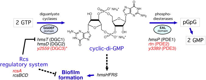
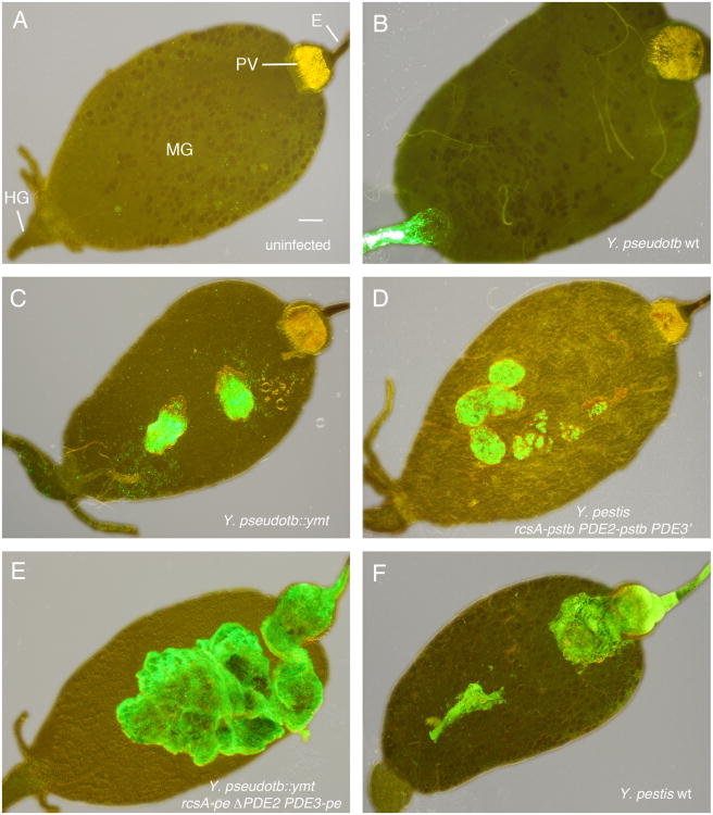
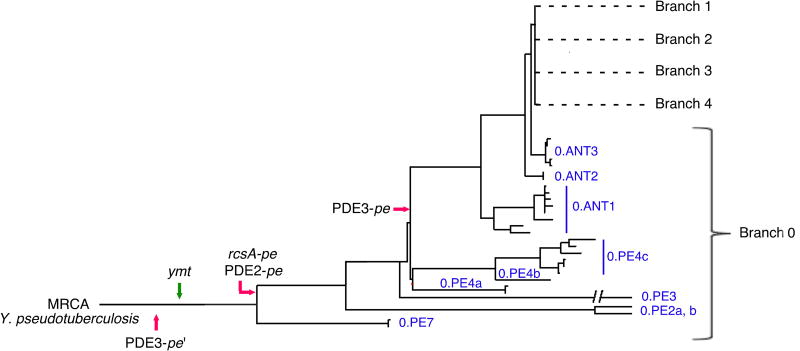
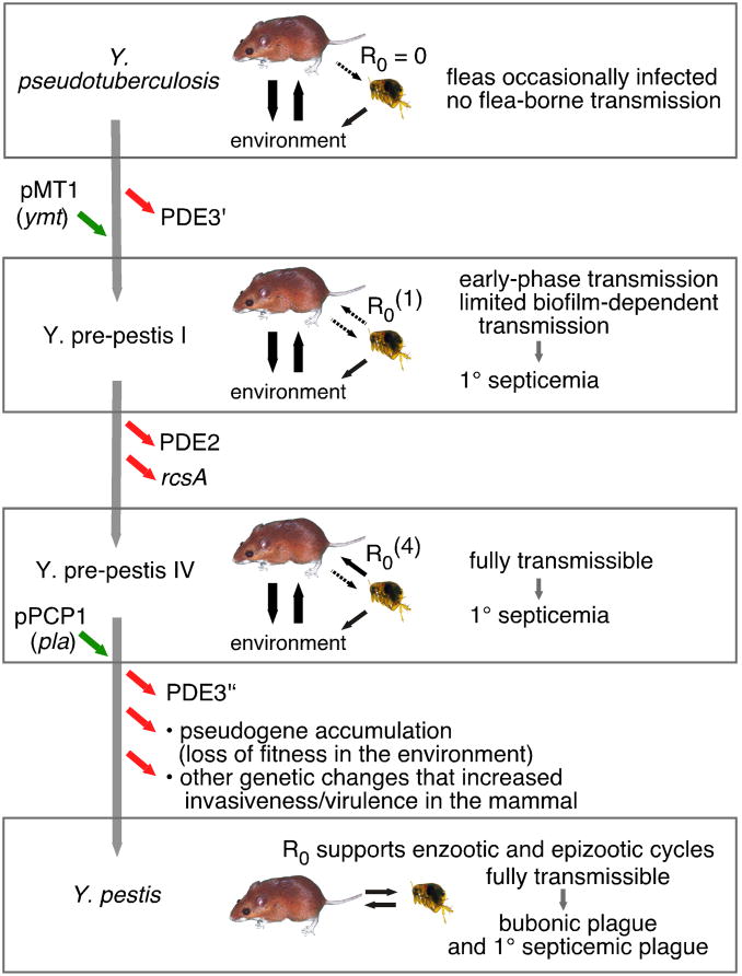
Yersinia pestis is an arthropod-borne bacterial pathogen that evolved recently from Yersinia pseudotuberculosis, an enteric pathogen transmitted via the fecal-oral route. This radical ecological transition can be attributed to a few discrete genetic changes from a still-extant recent ancestor, thus providing a tractable case study in pathogen evolution and emergence. Here, we determined the genetic and mechanistic basis of the evolutionary adaptation of Y. pestis to flea-borne transmission. Remarkably, only four minor changes in the bacterial progenitor, representing one gene gain and three gene losses, enabled transmission by flea vectors. All three loss-of-function mutations enhanced cyclic-di-GMP-mediated bacterial biofilm formation in the flea foregut, which greatly increased transmissibility. Our results suggest a step-wise evolutionary model in which Y. pestis emerged as a flea-borne clone, with each genetic change incrementally reinforcing the transmission cycle. The model conforms well to the ecological theory of adaptive radiation.


Figures

Figure 1
Figure 1. Comparison of Genes Involved in c-di-GMP Metabolism and Biofilm Formation in Y. pseudotuberculosis and Y. pestis
Genes indicated in red are pseudogenes in Y. pestis KIM. *The DGC3 gene is functional in most Y. pestis strains. Dashed lines indicate indirect (and undefined) induction or repression mechanisms.
Figure 2
Figure 2. Effect of Four Genetic Changes on the Flea Infection Phenotype of Y. pestis and Y. pseudotuberculosis
Digestive tracts dissected from an uninfected flea (A) and from fleas 1-3 weeks after infection with the Y. pseudotuberculosis or Y. pestis strains indicated (B-F). All bacteria strains expressed green fluorescent protein and images were taken using combined phase contrast and fluorescent microscopy. Strain descriptions are in Table S1; -pstb indicates the Y. pseudotbuerculosis functional allele and -pe the Y. pestis pseudogene allele. E, esophagus; PV, proventriculus; MG, midgut; HG, hindgut. Scale bar = 0.1 mm.
Figure 3
Figure 3. Restoration of Three Y. pestis Pseudogenes Eliminates the Ability to Produce Proventricular Blockage in Fleas
(A) Percentages of fleas that developed proventricular blockage during the 4-wk period after feeding on blood containing the Y. pestis or Y. pseudotuberculosis strain indicated. (B, C) Percentages of fleas still infected (B) and average bacterial load per infected flea (C) 4 wks after the infectious blood meal. The PDE2-pstbE509A allele has a single nucleotide site-specific mutation in the EAL catalytic domain that eliminates enzyme activity, other allele notations are as in Figure 1 and Table S1. The mean and SEM (A, B) or range (C) of three independent experiments are shown; data for the Y. pestis rcsA-pstb strain are from (Sun et al., 2008). *, P < 0.0001 by Fisher's exact test; **, P = 0.007 and ***, P = 0.003 by Kruskal-Wallis test with Dunn's multiple comparison post-test.
Figure 4
Figure 4. Four Genetic Changes Enable Transmissibility by Fleabite
The number of CFU transmitted by fleas infected with the bacteria indicated was quantified at weekly intervals after a single infectious blood meal. Day 3 represents biofilm-independent early-phase transmission (EPT); later timepoints represent proventricular biofilm-dependent transmission. Numbers in parentheses associated with data points are the number of fleas that fed, and of those, the number that were completely or partially blocked. The maximum extent of proventricular infection achieved by the strains is shown at right.
Figure 5
Figure 5. Early Occurrence and Fixation of Genetic Changes that Enabled Flea-borne Transmission During the Emergence of Y. pestis
The earliest documented appearance in available genome sequences of gain of function (green arrow) and loss-of-function (red arrows) gene changes in the genealogy of Y. pestis is indicated. All changes are present in the Pestoides (0.PE) group in the root Branch 0 that are most closely related to the most recent common ancestor (MRCA), Y. pseudotuberculosis. The four changes in Y. pestis KIM are found in 0.ANT as well as all of the evolutionarily more recent Branch 1 and 2 strains. Genome sequences of the following were available for this analysis: Branch 0 strains 0.PE2a (Pestoides F), 0.PE3a (Angola), 0.PE4b (Pestoides A), 0.PE4c (91001), 0.ANT2a (B42003004) [The 0.PE7 strain contains ymt but the rcsA, PDE2, and PDE3 sequences were not accessible]; Branch 1 strains 1.ANTa (Antiqua), 1.ANT1b (UG05), 1.ANT1b (CP000308), 1.IN3g (E1979001), 1.ORI1 (A1122), 1.ORI3c (IP275), 1.ORI1d (CA88), 1.ORI1e (CO92), 1.ORI1g (FV-1), 1.ORI2u (F1991016), 1.ORI3b (MG05); Branch 2 strains 2.ANT1c (Nepal), 2.MED1a (KIM), 2.MED2c (K1973002), and 9 other unclassified biovar Orientalis strains. The figure and nomenclature are adapted from (Cui et al., 2013).
Figure 6
Figure 6. Model of the Evolutionary Route to Flea-borne Transmission of Y. pestis
A sequence of genetic changes and their incremental effect on transmissibility is diagramed (see text). Black arrows indicate transmission of bacteria between rodent, flea, and the external environment and their weight indicates ecological importance; dashed arrows indicate occasional stochastic transmission. Green arrows, gene gains; red arrows, gene losses. PDE3′ refers to the gene with the promoter region point mutation; PDE3″ to the gene with both the point and stop codon mutations (cf. Table 1 and Figure S3). pMT1 and pPCP1 refer to the two Y. pestis-specific plasmids that encode ymt and pla. The informal Y. pre-pestis nomenclature is adopted from (Carniel, 2003).

References

    1. Achtman M, Zurth K, Morelli G, Torrea G, Guiyoule A, Carniel E. Yersinia pestis, the cause of plague, is a recently emerged clone of Yersinia pseudotuberculosis. Proc Natl Acad Sci USA. 1999;96:14043–14048. - PMC - PubMed
    1. André JB, Day T. The effect of disease life history on the evolutionary emergence of novel pathogens. Proc Royal Soc B. 2005;272:1949–1956. - PMC - PubMed
    1. Antia R, Regoes RR, Koella JC, Bergstrom CT. The role of evolution in the emergence of infectious diseases. Nature. 2003;426:658–661. - PMC - PubMed
    1. Arinaminpathy N, McLean AR. Evolution and emergence of novel human infections. Proc Royal Soc B. 2009;276:3937–3943. - PMC - PubMed
    1. Bacot AW. Further notes on the mechanism of the transmission of plague by fleas. J Hyg. 1915;14:774–776. - PMC - PubMed

Publication types

MeSH terms

Substances

LinkOut - more resources