. 2014 Jul 8;53(26):4250-60.
doi: 10.1021/bi5003508. Epub 2014 Jun 25.

Structure of a sedoheptulose 7-phosphate cyclase: ValA from Streptomyces hygroscopicus

Affiliations

Structure of a sedoheptulose 7-phosphate cyclase: ValA from Streptomyces hygroscopicus

Kelsey M Kean et al. Biochemistry. .

Erratum in

  • Biochemistry. 2014 Jul 8;53(26):4316

Abstract

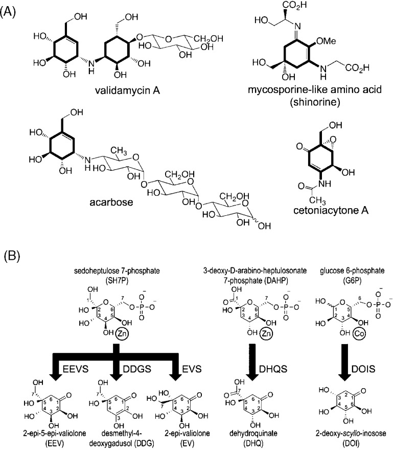
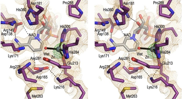
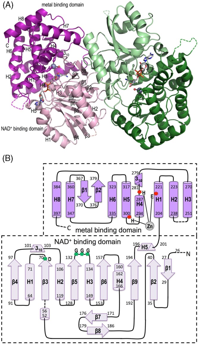
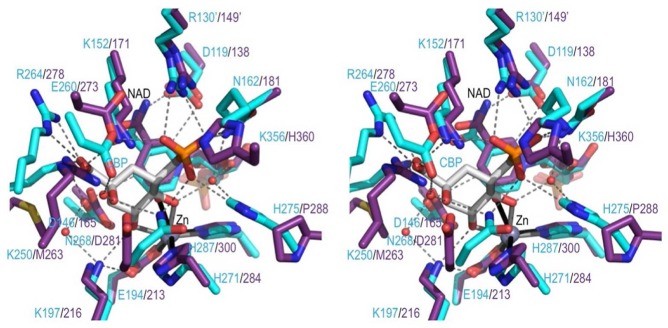
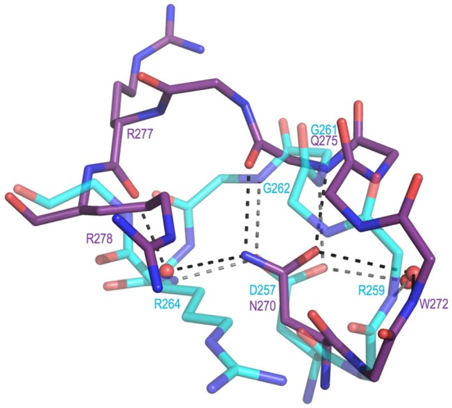
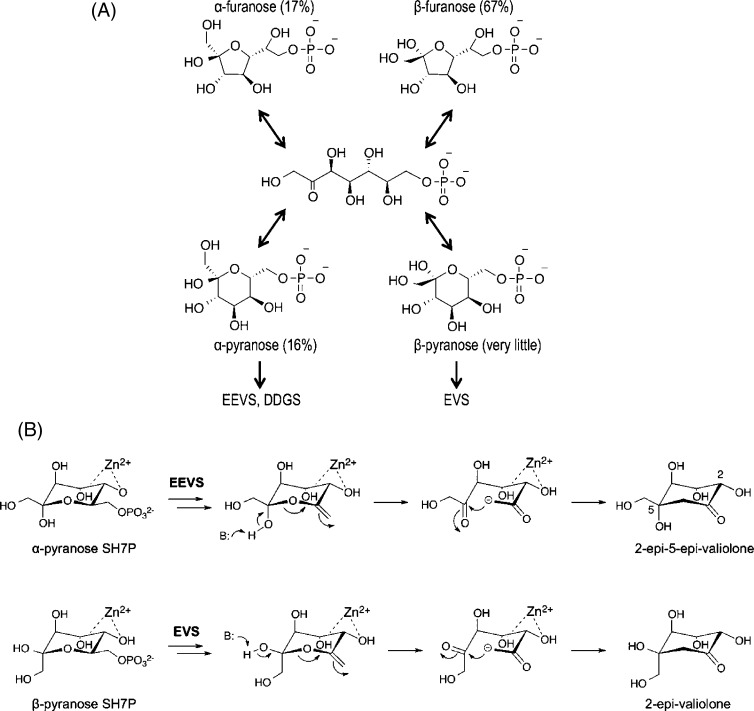
Sedoheptulose 7-phosphate cyclases (SH7PCs) encompass three enzymes involved in producing the core cyclitol structures of pseudoglycosides and similar bioactive natural products. One such enzyme is ValA from Streptomyces hygroscopicus subsp. jinggangensis 5008, which makes 2-epi-5-epi-valiolone as part of the biosynthesis of the agricultural antifungal agent validamycin A. We present, as the first SH7PC structure, the 2.1 Å resolution crystal structure of ValA in complex with NAD+ and Zn2+ cofactors. ValA has a fold and active site organization resembling those of the sugar phosphate cyclase dehydroquinate synthase (DHQS) and contains two notable, previously unrecognized interactions between NAD+ and Asp side chains conserved in all sugar phosphate cyclases that may influence catalysis. Because the domains of ValA adopt a nearly closed conformation even though no sugar substrate is present, comparisons with a ligand-bound DHQS provide a model for aspects of substrate binding. One striking active site difference is a loop that adopts a distinct conformation as a result of an Asp→Asn change with respect to DHQS and alters the identity and orientation of a key Arg residue. This and other active site differences in ValA are mostly localized to areas where the ValA substrate differs from that of DHQS. Sequence comparisons with a second SH7PC making a product with distinct stereochemistry lead us to postulate that the product stereochemistry of a given SH7PC is not the result of events taking place during catalysis but is accomplished by selective binding of either the α or β pyranose anomer of the substrate.


Figures

Figure 1
Figure 1
Reactions catalyzed by known sugar phosphate cyclases. (A) Four cyclitol-containing natural products are shown and labeled by name, with their C7-cyclitol units made by SH7PCs highlighted in bold. (B) The substrates (above) and products (below) of five sugar phosphate cyclases are shown. A divalent metal cation is drawn next to the two substrate hydroxyls seen (for DHQS and DOIS) to coordinate it. SH7PCs and DHQS may utilize Zn2+ or Co2+ as the divalent metal cation, but are shown here with Zn2+, the metal present in this structure of ValA and in structures of AnDHQS. DOIS uses only Co2+ for catalysis and is depicted as such. Abbreviations for the substrates and products are introduced, with each enzyme abbreviation being that of its product followed by an additional S for “synthase”. Because of resonance, DDG has an internal symmetry so the stereoconfiguration at C5 of the product after it is released into solution is not uniquely defined. We draw it here with the same stereochemistry as EEV, anticipating the proposal we make in this work that the cyclization products of EEVS and DDGS have the same C5 configuration.
Figure 2
Figure 2
Electron density map quality and active site structure. Stereoview of the ValA active site residues (purple carbons) and a water (red sphere) that are near the NAD+ (gray carbons) and the Zn2+ (silver sphere) cofactors. Coordination bonds (black lines) and select H-bonds (black dashes) are shown along with the 2FoFc electron density (orange, contoured at 1ρrms) and an anomalous difference map (green, contoured at 12ρrms).
Figure 3
Figure 3
Overall structure and topology of ValA. (A) Ribbon diagrams of the two chains of the ValA dimer are shown in purple and green tones, respectively, with the N-terminal NAD+-binding domains in light hues and the C-terminal metal-binding domains in dark hues. Dashed lines indicate internal unmodeled backbone segments. The NAD+ and the Zn2+ with its coordinating ligands are shown (colored as in Figure 1). Secondary structural elements in each domain of one monomer are labeled. (B) Topology diagram showing α-helices (cylinders), β-stands (arrows), 310-helices (triangular prisms), and π-helices (wider cylinder) with their first and last residues given. The minimal length α- and 310-helices (five and three residues, respectively) are left out of the family secondary structure nomenclature. The domains are colored light and dark purple as indicated, and helices (H) and strands (β) common to the SPCs are named sequentially within each domain. Dashed lines denote unmodeled backbone segments. The three Zn2+-binding residues (red asterisks) and the glycine-rich turn and acidic residues (green asterisks) important for NAD+ binding are indicated.
Figure 4
Figure 4
Sequence alignment of ValA with representative related enzymes. The sequence of ValA is listed first, and its secondary structure elements are schematically shown above the sequence. Other sequences in descending order are AvDDGS (A. variabilis DDGS, Ava_3858), AmEVS (Ac. mirum EVS, Amir_2000), PDB entry 1DQS (As. nidulans DHQS), PDB entry 2D2X (B. circulans DOIS), and PDB entry 1JQ5 (B. stearothermophilus glycerol dehydrogenase). For the structurally known proteins, the residues in β-strands (yellow), α-helices (teal), 310-helices (blue), and π-helices (orange) are highlighted. Residues involved in metal binding (m), NAD+ binding (n), and substrate binding and/or catalysis (∗) are denoted below the sequences, and active site residues with notable variation (↓) are denoted above the sequences.
Figure 5
Figure 5
ValA overlay with closed and open DHQS structures. Shown are ribbon diagrams of an unliganded, open DHQS (blue, PDB entry 1NRX), a CBP-bound, closed DHQS (cyan, PDB entry 1DQS), and ValA (purple), all overlaid on the basis of their NAD+-binding domains. Pale hues are used for the NAD+-binding domains and NAD+ and dark hues for the metal-binding domains. For the sake of clarity, only select secondary structure elements of the metal-binding domain are shown along with the three zinc-ligating residues (sticks) and the zinc (gray sphere). The active site side chain that does not align well between DHQS (Arg264) and ValA (Arg277) is colored green for both structures, and the alternate equivalent ValA residue (Arg278) is colored orange. The fact that other open DHQS structures, such as those of TtDHQS (PDB entry 1UJN) and HpDHQS (PDB entry 3CLH), are up to 5° different in domain orientation compared with AnDHQS (PDB entry 1NRX) does not alter the conclusions of this analysis.
Figure 6
Figure 6
Comparing the ValA active site region with the DHQS·CBP complex. Stereoview of select active site residues in ValA (purple) overlaid on the DHQS (cyan) in complex with CBP (white) shown in roughly the same orientation as DAHP in Figure 1. H-Bonding interactions in the DHQS active site (dashed lines) and coordination bonds with Zn2+ (solid lines) are shown. A prime on a residue number means it is from the other subunit of the dimer.
Figure 7
Figure 7
Active site loop difference that relates to the presence of Asp257 in DHQS vs Asn270 in ValA. Shown are residues 269–278 of ValA (purple) and residues 256–264 of DHQS (cyan, PDB entry 1DQS) after the proteins have been overlaid as in Figure 5. H-Bonding interactions (dashed lines) involving the loop residues and associated waters are shown. In DHQS, the Asp257 carboxylate receives H-bonds directly or indirectly (via water) from four backbone nitrogens (from Arg259, Gly261, Gly262, and Arg264). In ValA, the Asn270 side chain amide directly or indirectly makes H-bonds with two backbone nitrogens (from Trp272 and Glu275) and two backbone oxygens (from Gln275 and Arg277).
Figure 8
Figure 8
Variation in active site residues among sugar phosphate cyclases. Schematic drawing of residues lining the substrate-binding pocket in the DHQS·CBP complex shown in roughly the same orientation as in Figure 6. Each DHQS residue shown is labeled (cyan), and under that label are listed the corresponding residues found in the structures of DOIS (pink) and EEVS (purple) or, on the basis of the alignment in Figure 4, residues expected to be equivalent in DDGS and EVS (black). The CBP ligand is shown in bold. H-Bond interactions (dashed lines) and coordination bonds with Zn2+ (solid lines) are shown. The residue numbering corresponds to the representative proteins used in Figure 4 (AnDHQS, BcDOIS, ValA, AvDDGS, and AmEVS).
Figure 9
Figure 9
Proposed enzyme-specific selection of forms of sedoheptulose 7-phosphate. (A) The pyranose and furanose α- and β-anomers of SH7P with their relative abundance as determined by NMR are shown along with the linear form via which they interconvert. Also shown is our proposal that EEVS and DDGS bind the α-pyranose anomer while EVS binds the β-pyranose anomer. (B) Ring opening and intramolecular aldol condensation steps of the proposed reaction mechanisms of EEVS and EVS, emphasizing how the α- and β-anomers of pyranose of SH7P are preorganized for the generation of the respective stereochemistries at C5 in the products. B represents an active site base that may aid ring opening.

References

    1. Mahmud T. (2003) The C7N aminocyclitol family of natural products. Nat. Prod. Rep. 20, 137–166. - PubMed
    1. Stratmann A.; Mahmud T.; Lee S.; Distler J.; Floss H. G.; Piepersberg W. (1999) The AcbC protein from Actinoplanes species is a C-7-cyclitol synthase related to 3-dehydroquinate synthases and is involved in the biosynthesis of the α-glucosidase inhibitor acarbose. J. Biol. Chem. 274, 10889–10896. - PubMed
    1. Yu Y.; Bai L. Q.; Minagawa K.; Jian X. H.; Li L.; Li J. L.; Chen S. Y.; Cao E. H.; Mahmud T.; Floss H. G.; Zhou X. F.; Deng Z. X. (2005) Gene cluster responsible for validamycin biosynthesis in Streptomyces hygroscopicus subsp jinggangensis 5008. Appl. Environ. Microbiol. 71, 5066–5076. - PMC - PubMed
    1. Carpenter E. P.; Hawkins A. R.; Frost J. W.; Brown K. A. (1998) Structure of dehydroquinate synthase reveals an active site capable of multistep catalysis. Nature 394, 299–302. - PubMed
    1. Nango E.; Kumasaka T.; Hirayama T.; Tanaka N.; Eguchi T. (2008) Structure of 2-deoxy-scyllo-inosose synthase, a key enzyme in the biosynthesis of 2-deoxystreptamine-containing aminoglycoside antibiotics, in complex with a mechanism-based inhibitor and NAD+. Proteins: Struct., Funct., Bioinf. 70, 517–527. - PubMed

Publication types

MeSH terms

Associated data

LinkOut - more resources