. 2014 Aug;24(8):1735-41.
doi: 10.1007/s00330-014-3200-3. Epub 2014 May 16.

Thrombus imaging in acute stroke: correlation of thrombus length on susceptibility-weighted imaging with endovascular reperfusion success

Affiliations

Thrombus imaging in acute stroke: correlation of thrombus length on susceptibility-weighted imaging with endovascular reperfusion success

Christian Weisstanner et al. Eur Radiol. 2014 Aug.

Abstract

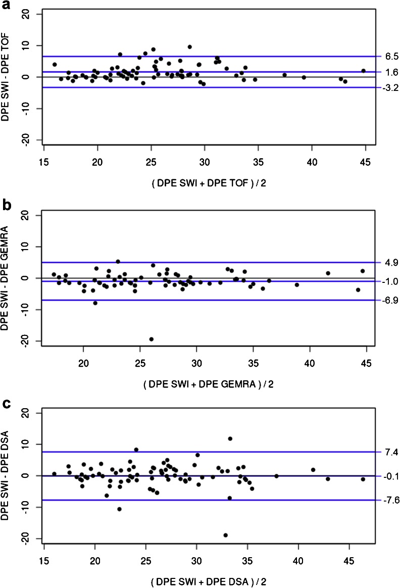
Objectives: Susceptibility-weighted imaging (SWI) enables visualization of thrombotic material in acute ischemic stroke. We aimed to validate the accuracy of thrombus depiction on SWI compared to time-of-flight MRA (TOF-MRA), first-pass gadolinium-enhanced MRA (GE-MRA) and digital subtraction angiography (DSA). Furthermore, we analysed the impact of thrombus length on reperfusion success with endovascular therapy.

Methods: Consecutive patients with acute ischemic stroke due to middle cerebral artery (MCA) occlusions undergoing endovascular recanalization were screened. Only patients with a pretreatment SWI were included. Thrombus visibility and location on SWI were compared to those on TOF-MRA, GE-MRA and DSA. The association between thrombus length on SWI and reperfusion success was studied.

Results: Eighty-four of the 88 patients included (95.5%) showed an MCA thrombus on SWI. Strong correlations between thrombus location on SWI and that on TOF-MRA (Pearson's correlation coefficient 0.918, P < 0.001), GE-MRA (0.887, P < 0.001) and DSA (0.841, P < 0.001) were observed. Successful reperfusion was not significantly related to thrombus length on SWI (P = 0.153; binary logistic regression).

Conclusions: In MCA occlusion thrombus location as seen on SWI correlates well with angiographic findings. In contrast to intravenous thrombolysis, thrombus length appears to have no impact on reperfusion success of endovascular therapy.

Key points: • SWI helps in assessing location and length of thrombi in the MCA • SWI, MRA and DSA are equivalent in detecting the MCA occlusion site • SWI is superior in identifying the distal end of the thrombus • Stent retrievers should be deployed over the distal thrombus end • Thrombus length did not affect success of endovascular reperfusion guided by SWI.


Figures

Fig. 1
Fig. 1
Measurement of thrombus localization. Midline was defined as a line connecting the occipital part of the superior sagittal sinus with the point midway between the A2 segments of the anterior cerebral arteries as seen on axial SWI and TOF-MRA. On coronal GE-MRA and anteroposterior DSA projections, midline was defined as a perpendicular line, midway between the A2 segments (OT occluding thrombus, IC infarct core, DPE distance to proximal end of thrombus, SSS superior sagittal sinus, ICA internal carotid artery, MCA middle cerebral artery, A2 A2 segment of the anterior cerebral artery)
Fig. 2
Fig. 2
60-year-old man with global aphasia and right-sided hemiparesis (NIHSS score 17). a On SWI a thrombus is visible in the left MCA. The distance of the proximal thrombus end to the midline on SWI is within the 1-mm range compared to that measured on b TOF-MRA, c GE-MRA and d DSA
Fig. 3
Fig. 3
Bland–Altman plots for measurements of distance of proximal thrombus end to midline (DPE) on SWI and corresponding measurements on TOF-MRA (a), GE-MRA (b) and DSA (c) showing mean bias and upper and lower limits of agreement (mean bias ± 1.96 × standard deviations). Units are in millimetres
Fig. 4
Fig. 4
Evaluation of thrombus anatomy (white arrows) on SWI (mIP). Patients with thrombi measuring less than 8 mm in length are preferentially treated with intravenous thrombolysis (a). Thrombi limited to the M1 segment but measuring more than 8 mm can usually be removed easily by stent retriever thrombectomy (b). Treatment of thrombotic occlusions extending from the M1 segment to one (c) or multiple (d) distal branches is generally more challenging. If the interventionist decides to deploy a stent retriever first a microcatheter has to be navigated blindly through the occluded vessel. For planning of this manoeuvre, SWI proves to be helpful as it visualizes the curvature of the occluded vessel segment

References

    1. Rha JH, Saver JL. The impact of recanalization on ischemic stroke outcome: a meta-analysis. Stroke. 2007;38:967–973. doi: 10.1161/01.STR.0000258112.14918.24. - DOI - PubMed
    1. Riedel CH, Zimmermann P, Jensen-Kondering U, Stingele R, Deuschl G, Jansen O. The importance of size: successful recanalization by intravenous thrombolysis in acute anterior stroke depends on thrombus length. Stroke. 2011;42:1775–1777. doi: 10.1161/STROKEAHA.110.609693. - DOI - PubMed
    1. Huang P, Chen CH, Lin WC, et al. Clinical applications of susceptibility weighted imaging in patients with major stroke. J Neurol. 2012;259:1426–1432. doi: 10.1007/s00415-011-6369-2. - DOI - PubMed
    1. Haacke EM, Mittal S, Wu Z, Neelavalli J, Cheng YC. Susceptibility-weighted imaging: technical aspects and clinical applications, part 1. AJNR Am J Neuroradiol. 2009;30:19–30. doi: 10.3174/ajnr.A1400. - DOI - PMC - PubMed
    1. Kao HW, Tsai FY, Hasso AN. Predicting stroke evolution: comparison of susceptibility-weighted MR imaging with MR perfusion. Eur Radiol. 2012;22:1397–1403. doi: 10.1007/s00330-012-2387-4. - DOI - PubMed

MeSH terms