

Quantitative expression and localization of cysteine and aspartic proteases in human abdominal aortic aneurysms
- PMID: 24833013
- PMCID: PMC3972792
- DOI: 10.1038/emm.2014.20
Quantitative expression and localization of cysteine and aspartic proteases in human abdominal aortic aneurysms
Abstract
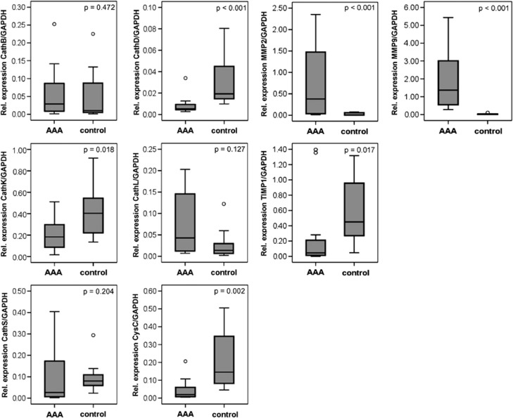
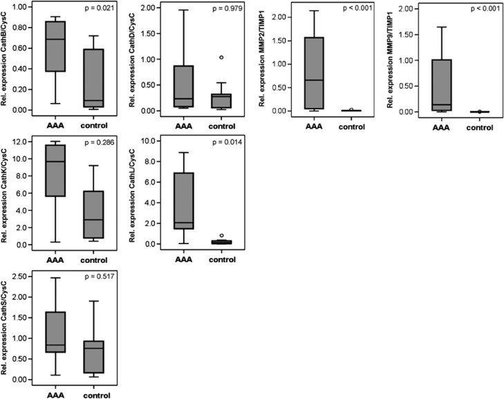
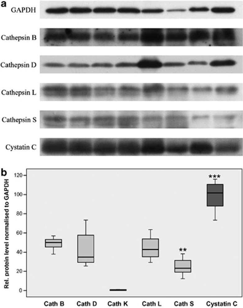
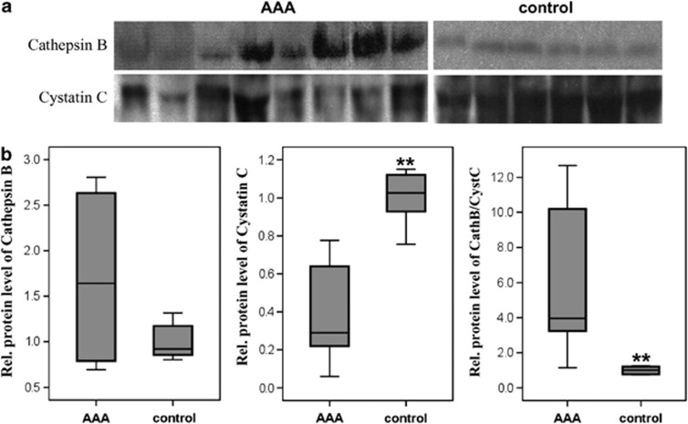
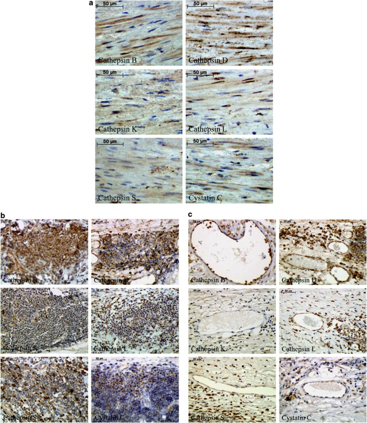
Cysteine and aspartic proteases possess high elastolytic activity and might contribute to the degradation of the abdominal aortic aneurysm (AAA) wall. The aim of this study was to analyze, in detail, the proteases (cathepsins B, D, K, L and S, and inhibitor cystatin C) found in human AAA and healthy aortic tissue samples. The vessel walls from AAA patients (n=36) and nonaneurysmal aortae (n=10) were retrieved using conventional surgical repair and autopsy methods. Serum samples from the same AAA patients and 10 healthy volunteers were also collected. Quantitative expression analyses were performed at the mRNA level using real-time reverse transcriptase-PCR (RT-PCR). Furthermore, analyses at the protein level included western blot and immunoprecipitation analyses. Cellular sources of cysteine/aspartic proteases and cystatin C were identified by immunohistochemistry (IHC). All cysteine/aspartic proteases and cystatin C were detected in the AAA and control samples. Using quantitative RT-PCR, a significant increase in expression was observed for cathepsins B (P=0.021) and L (P=0.018), compared with the controls. Cathepsin B and cystatin C were also detected in the serum of AAA patients. Using IHC, smooth muscle cells (SMCs) and macrophages were positive for all of the tested cathepsins, as well as cystatin C; in addition, the lymphocytes were mainly positive for cathepsin B, followed by cathepsins D and S. All cysteine/aspartic proteases analyzed in our study were detected in the AAA and healthy aorta. The highest expression was found in macrophages and SMCs. Consequently, cysteine/aspartic proteases might play a substantial role in AAA.
Figures
Similar articles
-
Cathepsin S is associated with degradation of collagen I in abdominal aortic aneurysm.Vasa. 2018 Jun;47(4):285-293. doi: 10.1024/0301-1526/a000701. Epub 2018 Apr 6. Vasa. 2018. PMID: 29624112
-
Cysteine protease activity in the wall of abdominal aortic aneurysms.J Vasc Surg. 2007 Dec;46(6):1260-6. doi: 10.1016/j.jvs.2007.08.015. J Vasc Surg. 2007. PMID: 18155003
-
Histopathological analysis of cellular localization of cathepsins in abdominal aortic aneurysm wall.Int J Exp Pathol. 2012 Aug;93(4):252-8. doi: 10.1111/j.1365-2613.2012.00819.x. Int J Exp Pathol. 2012. PMID: 22804761 Free PMC article.
-
Cysteine protease cathepsins and matrix metalloproteinases in the development of abdominal aortic aneurysms.Future Cardiol. 2013 Jan;9(1):89-103. doi: 10.2217/fca.12.71. Future Cardiol. 2013. PMID: 23259477 Free PMC article. Review.
-
Pathogenesis of abdominal aortic aneurysms: microRNAs, proteases, genetic associations.Annu Rev Med. 2014;65:49-62. doi: 10.1146/annurev-med-101712-174206. Epub 2013 Nov 21. Annu Rev Med. 2014. PMID: 24274177 Review.
Cited by
-
Abdominal aortic aneurysm: novel mechanisms and therapies.Curr Opin Cardiol. 2015 Nov;30(6):566-73. doi: 10.1097/HCO.0000000000000216. Curr Opin Cardiol. 2015. PMID: 26352243 Free PMC article. Review.
-
Plasma Cystatin B Association With Abdominal Aortic Aneurysms and Need for Later Surgical Repair: A Sub-study of the VIVA Trial.Eur J Vasc Endovasc Surg. 2018 Dec;56(6):826-832. doi: 10.1016/j.ejvs.2018.08.028. Epub 2018 Sep 24. Eur J Vasc Endovasc Surg. 2018. PMID: 30262158 Free PMC article.
-
Aortic Aneurysms.Arterioscler Thromb Vasc Biol. 2017 Jun;37(6):e59-e65. doi: 10.1161/ATVBAHA.117.309578. Arterioscler Thromb Vasc Biol. 2017. PMID: 28539494 Free PMC article. Review. No abstract available.
-
The role of vascular smooth muscle cells in the development of aortic aneurysms and dissections.Eur J Clin Invest. 2022 Apr;52(4):e13697. doi: 10.1111/eci.13697. Epub 2021 Nov 21. Eur J Clin Invest. 2022. PMID: 34698377 Free PMC article. Review.
-
Grape-seed Polyphenols Play a Protective Role in Elastase-induced Abdominal Aortic Aneurysm in Mice.Sci Rep. 2017 Aug 24;7(1):9402. doi: 10.1038/s41598-017-09674-4. Sci Rep. 2017. PMID: 28839206 Free PMC article.
References
-
- Reeps C, Pelisek J, Seidl S, Schuster T, Zimmermann A, Kuehnl A, et al. Inflammatory infiltrates and neovessels are relevant sources of MMPs in abdominal aortic aneurysm wall. Pathobiology. 2009;76:243–252. - PubMed
-
- Heider P, Wolf O, Reeps C, Hanke M, Zimmermann A, Berger H, et al. Aneurysms and dissections of the thoracal and abdominal aorta Chirurg 200778600,602–606, 608–610. - PubMed
-
- Shimizu K, Mitchell RN, Libby P. Inflammation and cellular immune responses in abdominal aortic aneurysms. Arterioscler Thromb Vasc Biol. 2006;26:987–994. - PubMed
-
- Brewster DC, Cronenwett JL, Hallett JW, Johnston KW, Krupski WC, Matsumura JS. Guidelines for the treatment of abdominal aortic aneurysms. Report of a subcommittee of the Joint Council of the American Association for Vascular Surgery and Society for Vascular Surgery. J Vasc Surg. 2003;37:1106–1117. - PubMed
MeSH terms
Substances
LinkOut - more resources
Full Text Sources
Other Literature Sources