

A rare case of anterior chest wall schwannoma masquerading as a breast tumor
- PMID: 24833138
- PMCID: PMC4027899
- DOI: 10.9738/INTSURG-D-13-00145.1
A rare case of anterior chest wall schwannoma masquerading as a breast tumor
Abstract
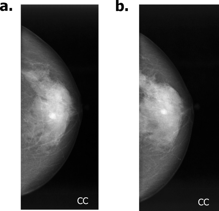
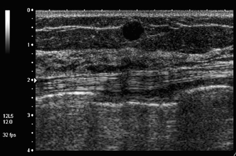
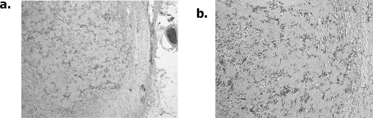
A schwannoma is a tumor that develops on peripheral nerves or spinal roots. Although any part of the body can be affected, the breast is a quite unusual site for schwannomas. We report herein a case of schwannoma presenting as a breast tumor. In the current case, the tumor showed both clinically and mammographically as a well-defined breast mass. Of interest, sonographically, the well-defined mass appeared to be located in subcutaneous tissue, not in breast parenchyma, and this finding was confirmed histopathologically. These findings indicate the possibility that a schwannoma arising from subcutaneous breast tissue can show exophytic growth to the breast and appear as a breast tumor. In other words, our case implies the possible presence of a "pseudo" breast schwannoma.
Keywords: Breast; Breast cancer; Schwannoma.
Figures
References
-
- Das Gupta TK, Brasfield RD, Strong EW, Hajdu SI. Benign solitary schwannomas (neurilemomas) Cancer. 1969;24(2):355–366. - PubMed
-
- Uchida N, Yokoo H, Kuwano H. Schwannoma of the breast: report of a case. Surg Today. 2005;35(3):238–242. - PubMed
-
- Bellezza G, Lombardi T, Panzarola P, Sidoni A, Cavaliere A, Giansanti M. Schwannoma of the breast: a case report and review of the literature. Tumori. 2007;93(3):308–311. - PubMed
-
- Balci P, Pekcevik YT, Caferova S, Canda T, Sevinc A, Saydam S. A case of benign schwannoma of the breast: mammographic, ultrasonographic and color Doppler ultrasonographic findings. Breast J. 2009;15(4):417–418. - PubMed
Publication types
MeSH terms
LinkOut - more resources
Full Text Sources
Other Literature Sources
Medical
