. 2014 Oct;15(5):592-600.
doi: 10.1089/sur.2013.016. Epub 2014 May 15.

Bacterial biofilms on implanted suture material are a cause of surgical site infection

Affiliations

Bacterial biofilms on implanted suture material are a cause of surgical site infection

Sandeep Kathju et al. Surg Infect (Larchmt). 2014 Oct.

Abstract

Background: Surgical site infection (SSI) has been estimated to occur in up to 5% of all procedures, accounting for up to 0.5% of all hospital costs. Bacterial biofilms residing on implanted foreign bodies have been implicated as contributing or causative factors in a wide variety of infectious scenarios, but little consideration has been given to the potential for implanted, submerged suture material to act as a host for biofilm and thus serve as a nidus of infection.

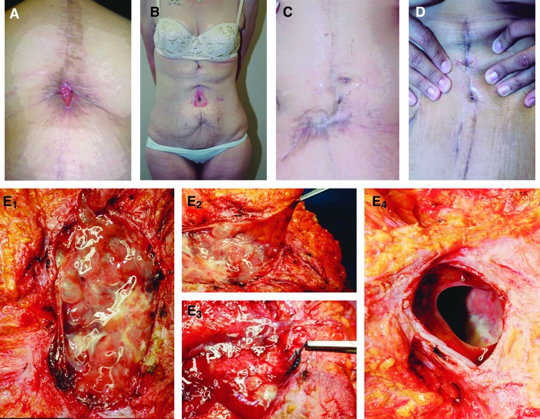
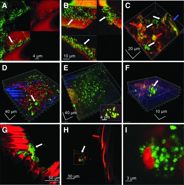
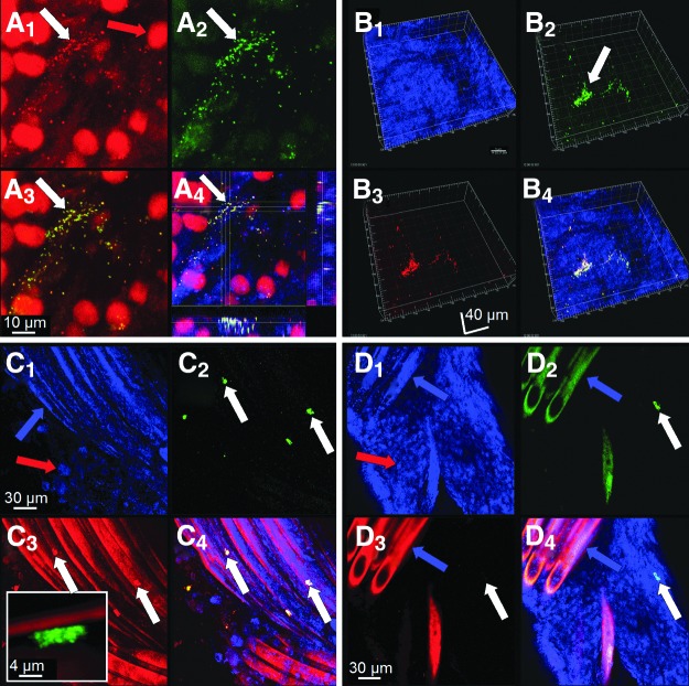
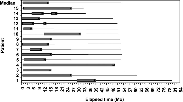
Methods: We report a series of 15 patients who underwent open Roux-en-Y gastric bypass (with musculofascial closure with permanent, multifilament sutures) who developed longstanding and refractory SSIs in the abdominal wall. Explanted suture material at subsequent exploration was examined for biofilm with confocal laser-scanning microscopy (CLSM) and fluorescence in situ hybridization (FISH).

Results: All 15 patients at re-exploration were found to have gross evidence of a "slimy" matrix or dense reactive granulation tissue localized to the implanted sutures. Confocal laser-scanning microscopy revealed abundant biofilm present on all sutures examined; FISH was able to identify the presence of specific pathogens in the biofilm. Complete removal of the foreign bodies (and attendant biofilms) resulted in all cases in cure of the SSI.

Conclusion: Bacterial biofilms on implanted suture material can manifest as persistent surgical site infections that require complete removal of the underlying foreign body substrata for resolution.


Figures

<b>FIG. 1.</b>
FIG. 1.
Pre- and intra-operative photographs of patients presenting with chronic surgical site infections after Roux-en-Y gastric bypass. A–D: Pre-operative views of multiple chronic SSIs with varying presentations. Note that the chronic wounds associated with the SSI could appear, among other things, granular (A), excoriated (B), involved with dense reactive scar (C), or unimpressive with just scant drainage (D). E1 – E4: “Slimy” accumulation consistent with biofilm infection situated on a mass of suture material in the abdominal wall. E2 depicts the viscosity of the material. E3 shows that this process is centered on embedded green polyester suture. E4 depicts the full-thickness muscle/fascia defect left after removal (by curettage) of sutures and associated infected tissues. Note the smooth, remodeled edges, indicating that chronic suture-associated SSI can be a cause of hernia formation. Color image is available online at www.liebertpub.com/sur
<b>FIG. 2.</b>
FIG. 2.
Confocal microscopic examination of suture and associated reactive tissue stained with the BacLight Live/Dead kit, showing various aspects of viable bacteria in biofilm configuration (indicated by white arrows). Images shown are representative from five patients. A) Biofilm consisting largely of cocci growing between the suture braids. The upper left panel shows live bacteria (green), and the upper right panel shows dead bacteria (red) and the autofluorescent sutures. The lower left panel is an overlay. B) Cross-section through the braids of a multifilament suture shows that the biofilm bacteria had penetrated the crevice formed by individual braids. The upper left panel is a maximum projection XY “plan” view and saggital sections in the XY and XZ planes are to the right and below respectively. The biofilm was up to 30 microns thick. C) The patient shown in panel B also had clusters of cocci in biofilms attached to the associated reactive tissue that had invested the suture material (blue arrow). The nuclei of host inflammatory cells were also seen (red arrow). D) Clusters of live coccal biofilm attached to the fibrous sheath (the nuclei of host cells are red) which was itself overlying the braids of the sutures (evident as blue striations). E) Extensive coccal biofilm attached to the fibrous sheath investing the suture. Inset is high magnification of a cluster of biofilm cocci. F) Biofilm clusters of live cocci attached to sclerotic fibrous tissue that was in intimate association with the suture. G) Clusters of viable bacteria attached to the suture braids (red). H) In some cases clusters of bacteria (white arrow) were in close vicinity to the sutures (red arrow) but not directly attached. This is consistent with the presence of EPS surrounding the bacterial cluster and attaching it to the suture surface. I) Higher magnification showed that the cluster in panel H was made up of live coccal cells (green) in close proximity to a host cell (red). Some of these bacteria appear to be in the process of division. Color image is available online at www.liebertpub.com/sur
<b>FIG. 3.</b>
FIG. 3.
A FISH examination of suture and associated reactive tissue from two patients. Specimens were stained with either 1) the general 16S rRNA probe Eub388, or 2) the nucleic acid stain propidium iodide and either the Staphylococcus genus probe (Sta) or the S. aureus specific probe (Sau). A) A1 shows clusters of cocci (white arrow) and the nuclei of host cells (red arrow). A2 shows that the bacteria were staphylococci (green). A3 shows an overlay of the previous panels, and A4 is a plan view and saggital sections through the staphylococcal biofilm cluster attached to fibrous tissue in this patient. In this last image, cytoskeletal elements of host cells were also visualized by staining f-actin blue with phalloidin. B–D) Specimens from a second patient stained for S. aureus (Sau). B1 shows the surface of reactive tissue visualized by reflected light. B2 shows the Eub388 eubacterial probe specifically staining bacteria in a biofilm cell cluster green (white arrow). B3 shows that the biofilm cluster was composed of S. aureus (in this case red). B4 is an overlay; the mixed red and green signals from the doubly-stained organisms appear yellow. C) S. aureus biofilm clusters adherent directly to the braids of a suture. C1 is a reflected image showing the suture braids (blue arrow) and some of the associated host cells (red arrow). C2 shows four clusters of biofilm stained with Eub388 (green). C3 shows that they were S. aureus (red). Inset shows a high magnification image of one cluster of cocci attached directly to the suture. C4 is an overlay. D) Evidence that the infection in this patient was polymicrobial. D1 is imaged using reflected light, showing suture braids and host cells. D2 shows a cluster of biofilm stained with the Eub388 probe (white arrow). However, the same cluster fails to stain with the Sau probe (D3), demonstrating that in addition to S. aureus the patient was infected with another species of bacteria. D4 is an overlay. Color images available online at www.liebertpub.com/sur
<b>FiG. 4.</b>
FiG. 4.
Patient timelines, from the time of open RYGB surgery to time of writing. Time “0” represents the RYGB surgery at which polyester sutures were placed. The initial solid line represents an initial disease-free condition. The appearance of an open gray bar signifies the presentation of the SSI. The length of the gray bar is the length of persisting SSI. The asterisk (*) indicates the panniculectomy and abdominal wall exploration surgery at which suture material was removed. The solid lines thereafter represent a disease-free condition again. In two patients, (as discussed in Results) two surgical interventions were undertaken, hence the appearance of two gray bars. All patients, after complete removal of suture material, have remained free of SSI.

Similar articles

Cited by

References

    1. Cheadle WG. Risk factors for surgical site infection. Surg Infect (Larchmt) 2006;7Suppl 1:S7–S11 - PubMed
    1. Fry DE. The economic costs of surgical site infection. Surg Infect (Larchmt) 2002;3Suppl 1:S37–S43 - PubMed
    1. Edmiston CE, Seabrook GR, Goheen MP, et al. . Bacterial adherence to surgical sutures: Can antibacterial-coated sutures reduce the risk of microbial contamination? J Am Coll Surg 2006;203:481–489 - PubMed
    1. Turina M, Cheadle WG. Management of established surgical site infections. Surg Infect (Larchmt) 2006; 7Suppl 3:S33–S41 - PubMed
    1. Rasnake MS, Dooley DP. Culture-negative surgical site infections. Surg Infect (Larchmt) 2006;7:555–565 - PubMed

Publication types

Substances

LinkOut - more resources