

An Approach to Breast Cancer Diagnosis via PET Imaging of Microcalcifications Using (18)F-NaF
- PMID: 24833491
- PMCID: PMC4320039
- DOI: 10.2967/jnumed.114.139170
An Approach to Breast Cancer Diagnosis via PET Imaging of Microcalcifications Using (18)F-NaF
Abstract
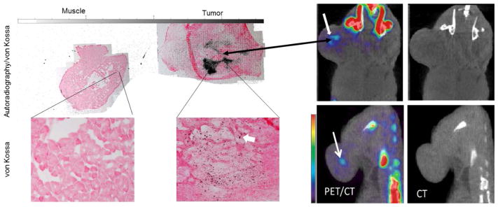
Current radiologic methods for diagnosing breast cancer detect specific morphologic features of solid tumors or any associated calcium deposits. These deposits originate from an early molecular microcalcification process of 2 types: type 1 is calcium oxylate and type II is carbonated calcium hydroxyapatite. Type I microcalcifications are associated mainly with benign tumors, whereas type II microcalcifications are produced internally by malignant cells. No current noninvasive in vivo techniques are available for detecting intratumoral microcalcifications. Such a technique would have a significant impact on breast cancer diagnosis and prognosis in preclinical and clinical settings. (18)F-NaF PET has been used solely for bone imaging by targeting the bone hydroxyapatite. In this work, we provide preliminary evidence that (18)F-NaF PET imaging can be used to detect breast cancer by targeting the hydroxyapatite lattice within the tumor microenvironment with high specificity and soft-tissue contrast-to-background ratio while delineating tumors from inflammation.
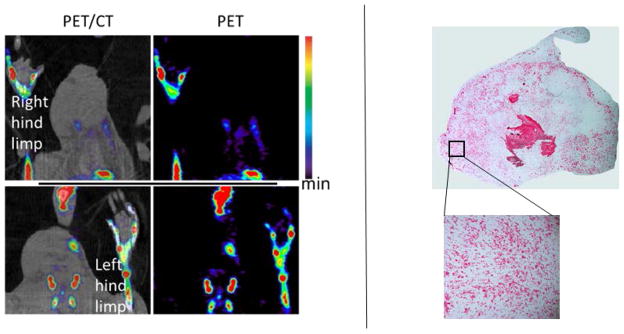
Methods: Mice were injected with approximately 10(6) MDA-MB-231 cells subcutaneously and imaged with (18)F-NaF PET/CT in a 120-min dynamic sequence when the tumors reached a size of 200-400 mm(3). Regions of interest were drawn around the tumor, muscle, and bone. The concentrations of radiotracer within those regions of interest were compared with one another. For comparison to inflammation, rats with inflamed paws were subjected to (18)F-NaF PET imaging.
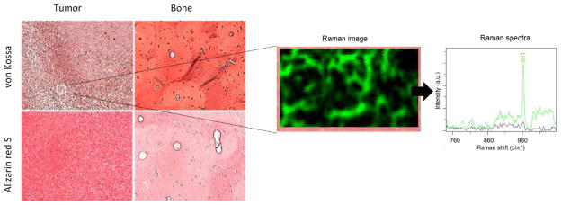
Results: Tumor uptake of (18)F(-) was significantly higher (P < 0.05) than muscle uptake, with the tumor-to-muscle ratio being about 3.5. The presence of type II microcalcification in the MDA-MB-231 cell line was confirmed histologically using alizarin red S and von Kossa staining as well as Raman microspectroscopy. No uptake of (18)F(-) was observed in the inflamed tissue of the rats. Lack of hydroxyapatite in the inflamed tissue was verified histologically.
Conclusion: This study provides preliminary evidence suggesting that specific targeting with (18)F(-) of hydroxyapatite within the tumor microenvironment may be able to distinguish between inflammation and cancer.
Keywords: 18F-NaF; 18F−; MDA-MB-231; PET; hydroxyapatite; breast cancer; calcium oxalate; microcalcification.
© 2014 by the Society of Nuclear Medicine and Molecular Imaging, Inc.
Figures
Similar articles
-
Detection of breast cancer microcalcification using (99m)Tc-MDP SPECT or Osteosense 750EX FMT imaging.Nucl Med Biol. 2015 Mar;42(3):269-73. doi: 10.1016/j.nucmedbio.2014.11.010. Epub 2014 Dec 6. Nucl Med Biol. 2015. PMID: 25533764 Free PMC article.
-
More advantages in detecting bone and soft tissue metastases from prostate cancer using 18F-PSMA PET/CT.Hell J Nucl Med. 2019 Jan-Apr;22(1):6-9. doi: 10.1967/s002449910952. Epub 2019 Mar 7. Hell J Nucl Med. 2019. PMID: 30843003
-
Prospective Comparison of 99mTc-MDP Scintigraphy, Combined 18F-NaF and 18F-FDG PET/CT, and Whole-Body MRI in Patients with Breast and Prostate Cancer.J Nucl Med. 2015 Dec;56(12):1862-8. doi: 10.2967/jnumed.115.162610. Epub 2015 Sep 24. J Nucl Med. 2015. PMID: 26405167
-
The Role of 18F-Sodium Fluoride PET/CT Bone Scans in the Diagnosis of Metastatic Bone Disease from Breast and Prostate Cancer.J Nucl Med Technol. 2016 Dec;44(4):217-222. doi: 10.2967/jnmt.116.176859. Epub 2016 Sep 15. J Nucl Med Technol. 2016. PMID: 27634981 Review.
-
Newer PET application with an old tracer: role of 18F-NaF skeletal PET/CT in oncologic practice.Radiographics. 2014 Sep-Oct;34(5):1295-316. doi: 10.1148/rg.345130061. Radiographics. 2014. PMID: 25208282 Review.
Cited by
-
Hydroxyapatite Nanoparticles for Improved Cancer Theranostics.J Funct Biomater. 2022 Jul 20;13(3):100. doi: 10.3390/jfb13030100. J Funct Biomater. 2022. PMID: 35893468 Free PMC article. Review.
-
Detection of breast cancer microcalcification using (99m)Tc-MDP SPECT or Osteosense 750EX FMT imaging.Nucl Med Biol. 2015 Mar;42(3):269-73. doi: 10.1016/j.nucmedbio.2014.11.010. Epub 2014 Dec 6. Nucl Med Biol. 2015. PMID: 25533764 Free PMC article.
-
Breast Tumor Microcalcification Induced by Bone Morphogenetic Protein-2: A New Murine Model for Human Breast Tumor Diagnosis.Contrast Media Mol Imaging. 2018 Nov 11;2018:2082154. doi: 10.1155/2018/2082154. eCollection 2018. Contrast Media Mol Imaging. 2018. PMID: 30534026 Free PMC article.
-
Dicarbonyl Electrophiles Mediate Inflammation-Induced Gastrointestinal Carcinogenesis.Gastroenterology. 2021 Mar;160(4):1256-1268.e9. doi: 10.1053/j.gastro.2020.11.006. Epub 2020 Nov 13. Gastroenterology. 2021. PMID: 33189701 Free PMC article.
-
Current Approaches of Nuclear Molecular Imaging in Breast Cancer.Cancers (Basel). 2025 Jun 23;17(13):2105. doi: 10.3390/cancers17132105. Cancers (Basel). 2025. PMID: 40647404 Free PMC article. Review.
References
-
- DeSantis C, Siegel R, Bandi P, Jemal A. Breast cancer statistics, 2011. CA: a cancer journal for clinicians. 2011 Nov-Dec;61(6):409–418. - PubMed
-
- Agency for health care research and quality. Comparative Effectiveness of Non-Invasive Diagnostic Tests for Breast Abnormalities – An Update of a 2006 Report. Effective Health Care Program: Research Review. 2012 Feb
-
- Gershon-Cohen J. The importance of x-ray microcalcifications in breast cancer. The American journal of roentgenology, radium therapy, and nuclear medicine. 1967 Apr;99(4):1010–1011. - PubMed
-
- Going JJ, Anderson TJ, Crocker PR, Levison DA. Weddellite calcification in the breast: eighteen cases with implications for breast cancer screening. Histopathology. 1990 Feb;16(2):119–124. - PubMed
Publication types
MeSH terms
Substances
Grants and funding
LinkOut - more resources
Full Text Sources
Other Literature Sources
Medical
Miscellaneous