. 2014 Jul;3(7):821-35.
doi: 10.5966/sctm.2013-0212. Epub 2014 May 15.

Stable enhanced green fluorescent protein expression after differentiation and transplantation of reporter human induced pluripotent stem cells generated by AAVS1 transcription activator-like effector nucleases

Affiliations

Stable enhanced green fluorescent protein expression after differentiation and transplantation of reporter human induced pluripotent stem cells generated by AAVS1 transcription activator-like effector nucleases

Yongquan Luo et al. Stem Cells Transl Med. 2014 Jul.

Abstract

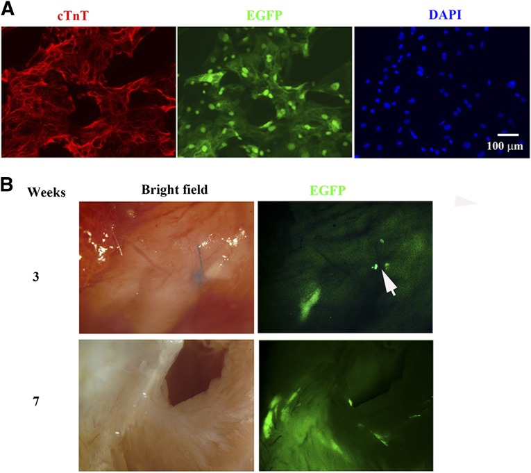
Human induced pluripotent stem (hiPS) cell lines with tissue-specific or ubiquitous reporter genes are extremely useful for optimizing in vitro differentiation conditions as well as for monitoring transplanted cells in vivo. The adeno-associated virus integration site 1 (AAVS1) locus has been used as a "safe harbor" locus for inserting transgenes because of its open chromatin structure, which permits transgene expression without insertional mutagenesis. However, it is not clear whether targeted transgene expression at the AAVS1 locus is always protected from silencing when driven by various promoters, especially after differentiation and transplantation from hiPS cells. In this paper, we describe a pair of transcription activator-like effector nucleases (TALENs) that enable more efficient genome editing than the commercially available zinc finger nuclease at the AAVS1 site. Using these TALENs for targeted gene addition, we find that the cytomegalovirus-immediate early enhancer/chicken β-actin/rabbit β-globin (CAG) promoter is better than cytomegalovirus 7 and elongation factor 1α short promoters in driving strong expression of the transgene. The two independent AAVS1, CAG, and enhanced green fluorescent protein (EGFP) hiPS cell reporter lines that we have developed do not show silencing of EGFP either in undifferentiated hiPS cells or in randomly and lineage-specifically differentiated cells or in teratomas. Transplanting cardiomyocytes from an engineered AAVS1-CAG-EGFP hiPS cell line in a myocardial infarcted mouse model showed persistent expression of the transgene for at least 7 weeks in vivo. Our results show that high-efficiency targeting can be obtained with open-source TALENs and that careful optimization of the reporter and transgene constructs results in stable and persistent expression in vitro and in vivo.

Keywords: AAVS1; Differentiation; Genome editing; Human induced pluripotent stem cells; Transcription activator-like effector nuclease (TALEN); Transplantation.


Figures

Figure 1.
Figure 1.
pZT-AAVS1 TALEN construction and EGFP rescue assay. (A): TALEN domains in the pZT vector contain shortened N- and C- termini from TALE13 and repeat variable residue units from the Golden Gate TALEN kit. (B): Scheme of the EGFP rescue assay for evaluating ZFN and TALEN activity. The sequences at the top are the AAVS1 TALEN target sequence and the AAVS1 ZFN target sequence. Bold fonts indicate binding sites. (C): Flow cytometry of 293T-EGIP*AAVS1 rescue at day 3. One microgram of tGFP donor and 5 μg of each ZFN or TALEN were used for transfection. The data are representative for at least three experiments. Abbreviations: EGFP, enhanced green fluorescent protein; TALEN, transcription activator-like effector nuclease; ZFN, zinc finger nuclease.
Figure 2.
Figure 2.
Nonhomologous-end joining (NHEJ) assay. (A): Specificity of the NHEJ assay in HEK293T cells. Five micrograms of control vector (“C” lanes) or 5 μg of each AAVS1 transcription activator-like effector nucleases (TALENs) vector (“T” lanes) was used per 106 cells in the transfection. (B): Time course. The HEK293T cells were nucleofected with or without 3 μg of each pZT-AAVS1 TALEN for different time periods, as indicated. (C): Concentration-dependent studies. The HEK293T cells were nucleofected with different amounts of pZT-AAVS1 TALENs from 0 to 5 μg of each in 106 cells. (D): Comparison of cutting activity between AAVS1 zinc finger nucleases and pZT-AAVS1 TALENs. The same amounts of plasmid (5 μg of each in 106 cells) were used for nucleofection. Abbreviations: bp, base pairs; C, control; FU, fluorescence unit; T, AAVS1 TALEN.
Figure 3.
Figure 3.
AAVS1 targeting. (A): A schematic illustration of AAVS1 gene targeting with the pAAVS1D-CMV.amilRFP-EF1α.copGFPpuro donor. amilRFP and copGFP were driven by cytomegalovirus 7 (CMV7) and elongation factor 1α promoters, respectively. 5′F/5′R and 3′F/3′R primers were used for 5′ and 3′ junction polymerase chain reaction (PCR) screening. The 5′ PCR junction is from intron 1 of the PPP1R12C gene, just upstream of the left homology arm (AAVS1-LA), to the CMV7 promoter of the donor, with a size of 1.2 kb. The 3′ PCR junction is from downstream of the right homology arm (AAVS1-RA) to the copGFP-T2A-puro-pA cassette of the donor with a size of 1.5 kb. The 5′pb is located in a region of left homology arm AAVS1-LA with a size of 704 bp; therefore, it can recognize WT, TI, and random integration. (B): Images of fluorescence of copGFP and amilRFP at days 1 and 2 after nucleofection of pZT-AAVS1 transcription activator-like effector nucleases and the donor in NCRM5 hiPS cells. Bar = 400 μm. (C): Shown are PCR screening results for selected clones. The bottom PCR products served as a genomic DNA template control used for the screening analysis. The + symbol indicates that nearly all cells are copGFP positive; the − symbol indicates that nearly all cells are copGFP negative; and the “+/−” symbol indicates that clones contain mixed copGFP-positive and -negative cells. (D): Non-AAVS1 targeted clones were further used to confirm copGFP and amilRFP mRNA expression by reverse transcriptase quantitative PCR. The data are presented as fold change relative to control parental NCRM5 human induced pluripotent stem cells. ∗p < .05 compared with corresponding parental NCRM5 cells (analysis of variance with Dunnett’s post hoc test). (E): Southern blot analysis. After SphI digestion, the 5′pb detects the WT allele as a 6.5-kb band and TI as a 3.8-kb band due to the introduction of an SphI site in the CMV promoter. Abbreviations: amilRFP, red fluorescent protein from Acropora millepora; bp, base pairs; copGFP, copepod green fluorescent protein; kb, kilobase pairs; 5′pb, 5′ (internal) probe; RFP, red fluorescent protein; TI, targeted integration; WT, wild type.
Figure 4.
Figure 4.
Both copGFP and amilRFP expression was silenced at the AAVS1 targeted site. (A): The pAAVS1D-CMV.amilRFP-EF1α.copGFPpuro targeted clone 8 was expanded and enriched for copGFP expression by reselection using puromycin. Cell that were copGFP negative were obtained using FACS from the same clone 8. Levels of expression of copGFP were measured using flow cytometry analysis. The cells were subject to Southern blot analysis using the same 5′ probe used in Figure 3, and Western blot was used for further confirmation of copGFP proteins. The mRNA levels of copGFP and amilRFP were measured using RT-qPCR and are shown as fold changes in corresponding control NCRM5 cells. ∗p < .05 compared with corresponding parental NCRM5 cells (analysis of variance with Dunnett’s post hoc test). (B): The top row shows AAVS1 gene targeting with the pAAVS1D-EF1α.copGFPpuro donor. NCRM5 cells were targeted with this donor in the presence of pZT-AAVS1 transcription activator-like effector nucleases. After puromycin selection, the clones were selected and confirmed to be integrated with Southern blot using the 5′ probe. Please note that the Southern blot hybridized targeting size is increased to 8.9 kb due to removal of the SphI site in this truncated pAAVS1D-EF1α.copGFPpuro construct. Expression of copGFP levels were measured using FACS analysis and further confirmed by Western blot. The copGFP mRNA levels were measured using RT-qPCR. ∗p < .05 compared with parental NCRM5 cells. (n = 3, Student’s t test). Abbreviations: amilRFP, red fluorescent protein from Acropora millepora; copGFP, copepod green fluorescent protein; FACS, fluorescence-activated cell sorting; 5′pb, 5′ (internal) probe; RI, random integration; RT-qPCR, reverse transcriptase quantitative polymerase chain reaction; TI, targeted integration; WT, wild type.
Figure 5.
Figure 5.
The chicken β actin (CAG) promoter drove EGFP expression at the AAVS1 locus. (A): The top rows show the pAAVS1-CAG-EGFP construct, targeting site, and location of the 5′ probe, which is the same as that used in Figure 3 for Southern blot analysis. Both ND2 and NCRM5 human induced pluripotent stem cell lines were targeted using this construct donor and pZT-AAVS1 transcription activator-like effector nucleases. The correctly targeted clones are indicated with red numbers. The common band between WT and TI in multiple clones indicates random integrations, which can be from different genome loci. Karyotype analyses were shown on the bottom. (B): Shown are images and fluorescence-activated cell sorting analysis of EGFP levels in targeted colonies. Scale bars = 400 μm. Abbreviations: c7, clone 7; c8, clone 8; EGFP, enhanced green fluorescent protein; 5′pb, 5′ (internal) probe; TI, targeted integration; WT, wild type.
Figure 6.
Figure 6.
Characterization of AAVS1-CAG-EGFP clones. All results shown are from the ND2-AAVS1-CAG-EGFP clone 7 (c7). (A): Immunocytochemical staining of the targeted colonies with pluripotent marker NANOG and TRA-1-60. (B): Immunostaining after 14 days of embryoid body (EB) spontaneous differentiation and direct differentiation into definitive endoderm. (C): Flow cytometry analyses of EGFP expression. Left, undifferentiated states at passage 13; right, 16 days after in vitro random differentiation via EB stage. (D): Xenografts within 6–7 weeks and histological images with hematoxylin and eosin staining. Shown are features in ectoderm, endoderm and mesoderm. Scale bars = 100 μm; arrow points to smooth muscle structure. (E): EGFP expression of the differentiated cells directly derived from the teratoma. Abbreviation: EGFP, enhanced green fluorescent protein.
Figure 7.
Figure 7.
The chicken β actin-driven EGFP expression at the AAVS1 locus is retained in long-term differentiated cardiomyocytes and in vivo engrafted mouse hearts. (A): EGFP expression still occurred in cardiomyocytes after 10 days of dissociation from beating cardiomyocytes derived from the ND2-AAVS1-CAG-EGFP line. Left, cTnT staining; right two images, EGFP and DAPI staining. (B): EGFP-expressed cardiomyocyte grafts in the infarcted mouse heart for different lengths of time. The arrow indicates ligation site of the left anterior descending coronary artery. Top row, mouse heart 3 weeks after transplantation; bottom row, cross-section of the left ventricle 7 weeks after transplantation. Abbreviations: cTnT, cardiac troponin T; DAPI, 4′,6-diamidino-2-phenylindole; EGFP, enhanced green fluorescent protein.

References

    1. Rao MS, Malik N. Assessing iPSC reprogramming methods for their suitability in translational medicine. J Cell Biochem. 2012;113:3061–3068. - PMC - PubMed
    1. Walia B, Satija N, Tripathi RP, et al. Induced pluripotent stem cells: Fundamentals and applications of the reprogramming process and its ramifications on regenerative medicine. Stem Cell Rev. 2012;8:100–115. - PubMed
    1. Deacon T, Dinsmore J, Costantini LC, et al. Blastula-stage stem cells can differentiate into dopaminergic and serotonergic neurons after transplantation. Exp Neurol. 1998;149:28–41. - PubMed
    1. Hentze H, Graichen R, Colman A. Cell therapy and the safety of embryonic stem cell-derived grafts. Trends Biotechnol. 2007;25:24–32. - PubMed
    1. Zou J, Maeder ML, Mali P, et al. Gene targeting of a disease-related gene in human induced pluripotent stem and embryonic stem cells. Cell Stem Cell. 2009;5:97–110. - PMC - PubMed

Publication types

MeSH terms