. 2014 Jul 18;115(3):354-63.
doi: 10.1161/CIRCRESAHA.115.303632. Epub 2014 May 15.

Cardiac-specific YAP activation improves cardiac function and survival in an experimental murine MI model

Affiliations

Cardiac-specific YAP activation improves cardiac function and survival in an experimental murine MI model

Zhiqiang Lin et al. Circ Res. .

Abstract

Rationale: Yes-associated protein (YAP), the terminal effector of the Hippo signaling pathway, is crucial for regulating embryonic cardiomyocyte proliferation.

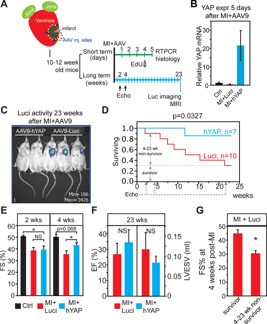
Objective: We hypothesized that YAP activation after myocardial infarction (MI) would preserve cardiac function and improve survival.

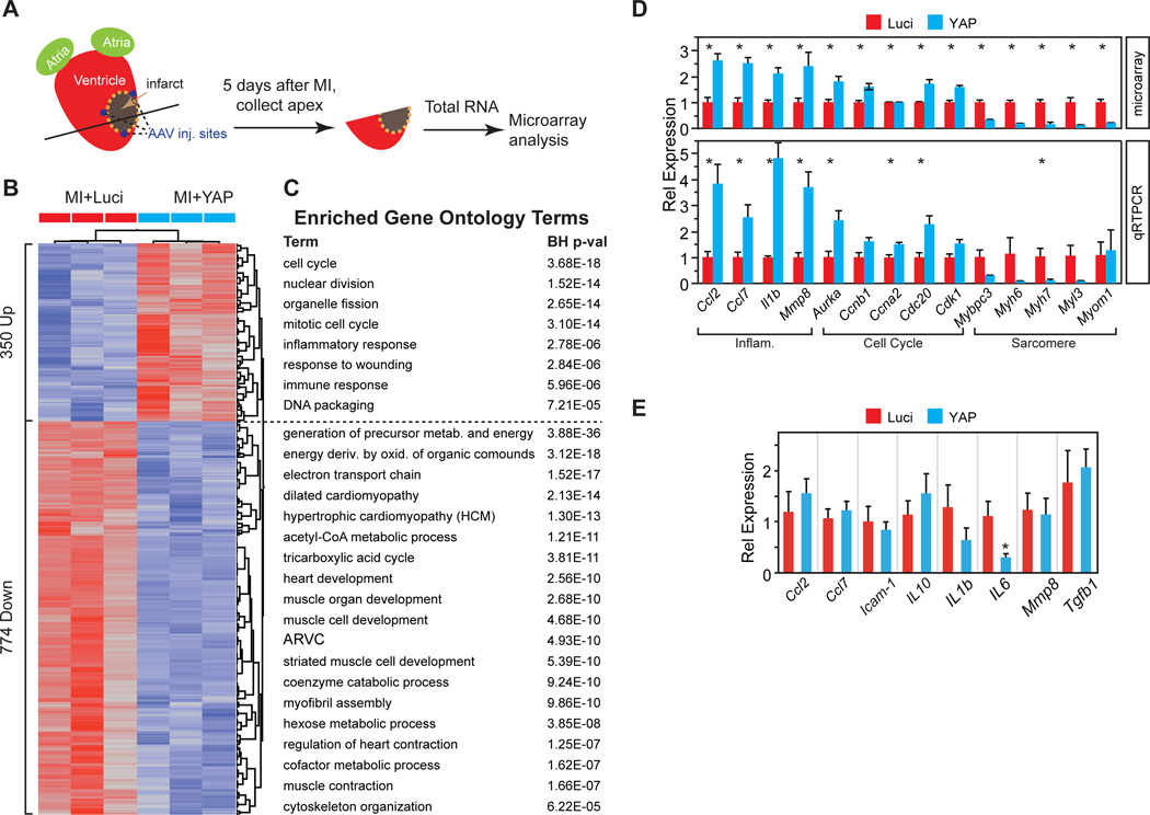
Methods and results: We used a cardiac-specific, inducible expression system to activate YAP in adult mouse heart. Activation of YAP in adult heart promoted cardiomyocyte proliferation and did not deleteriously affect heart function. Furthermore, YAP activation after MI preserved heart function and reduced infarct size. Using adeno-associated virus subtype 9 (AAV9) as a delivery vector, we expressed human YAP (hYAP) in the adult murine myocardium immediately after MI. We found that AAV9:hYAP significantly improved cardiac function and mouse survival. AAV9:hYAP did not exert its salutary effects by reducing cardiomyocyte apoptosis. Rather, AAV9:hYAP stimulated adult cardiomyocyte proliferation. Gene expression profiling indicated that AAV9:hYAP stimulated expression of cell cycle genes and promoted a less mature cardiac gene expression signature.

Conclusions: Cardiac-specific YAP activation after MI mitigated myocardial injury, improved cardiac function, and enhanced survival. These findings suggest that therapeutic activation of YAP or its downstream targets, potentially through AAV-mediated gene therapy, may be a strategy to improve outcome after MI.

Keywords: heart failure; myocardial infarction; regeneration; survival.


Conflict of interest statement

DISCLOSURES

The authors have no conflicts of interest to disclose.

Figures

Figure 1
Figure 1. Cardiac phenotype of YAPGOF mice
A. Dox-regulated, CM-specific expression system. B. Cardiac YAP expression of Dox-treated mice was measured by western blot. C. RT-PCR showing YAP overexpression in YAPGOF heart. D. Dox-dependent YAP expression in YAPGOF mice.
Figure 2
Figure 2. YAP stimulates adult CM proliferation
YAPGOF and control mice were treated with Dox from 4 to 8 weeks of life. Hearts were analyzed at 8 weeks. A. Heart weight to body weight ratio was not significantly different between groups. B–C. CM cross-sectional area was lower in YAPGOF than in control. WGA-stained sections were used to measure CM cross-sectional area. n=6–7. Bar = 25 µm. D. The relative number of CMs in YAPGOF and control hearts based on heart weight and CM cross-sectional area. *, P< 0.05. E–G. The size and number of CMs isolated from hearts by collagenase perfusion. n=4–8. Bar = 50 µm. H–I. The fraction of CMs bearing proliferative markers EdU or pH3 was determined in dissociated heart preparations. Yellow arrow indicates proliferating CM, and arrowhead indicates a non-myocyte. n=5. Bar = 25 µm. J. Nucleation of all CMs compared to EdU+ CMs was determined in dissociated cardiomyocytes. The large majority of proliferating CMs in control and YAPGOF groups were mononuclear.
Figure 3
Figure 3. Clonal analysis of YAP-induced adult CM proliferation
A–C. One color clonal analysis of CM proliferation. (A), Schematic of the experimental design. * indicates adjacent cells that were labeled by two separate recombination events, rather than by proliferation of a single labeled cell. (B), Representative images. Red arrows indicate a cluster of three red cells. (C), Quantification showing that clusters of 2 or more RFP+ cells were found more frequently in YAPGOF hearts. n=5 hearts per group. Bar = 50 µm or 10 µm. D–F. Two color clonal analysis of CM proliferation. (D), experimental design. * indicates a bichromatic cluster caused by two independent labeling events. (E), Representative images. Yellow arrows, bichromatic clusters. Red arrows, monochromatic clusters. The number of arrows sharing a common tail indicates the number of cells in that cluster. (F). Quantification showing that monochromatic clusters with ≥ 2 cells were more common in YAPGOF. Bichromatic clusters were not significantly different between groups. n=4 hearts per group. Bar = 25 µm.
Figure 4
Figure 4. Cardiac specific overexpression of YAP is well tolerated in adult mouse
Cardiac specific overexpression of YAP is well tolerated in adult mouse. A–B. 4 months of YAP induction in adult heart did not grossly affect heart size, morphology (A), or HW/BW ratio (B). A, bar=5 mm. C–D. Echocardiographic analysis of prolonged YAP overexpression on adult heart function. LVPW;d, diastolic left ventricular posterior wall thickness. *, P<0.05. n=4. E–F. Effect of YAP overexpression on myocardial fibrosis, assessed by picosirius red/fast green staining. (E), Representative sections. Fibrotic areas stain red. (F), Quantification. NS, not significant.
Figure 5
Figure 5. Activated YAP improved myocardial outcome after MI
A. Experimental design. B–C. MRI assessment of ventricular function. B, representative images. C, quantification. Bar = 5 mm. D. HW/BW was lower in YAPGOF. E–F. Scar size, measured as a percentage of ventricular circumference in sequential picro-sirus red and fast green stained sections. Bar = 5 mm. G. EdU+ CMs were more frequent in YAPGOF border and remote zones. Representative section shows an EdU+ CM (arrow) in YAPGOF. Bar = 25 µm. H. pH3+ CMs were more frequent in YAPGOF. Representative section shows a pH3+ CM (arrow). Bar = 10 µm.
Figure 6
Figure 6. Myocardial AAV9:hYAP injection post MI improved cardiac function and mouse survival
A. Schematic of AAV9:hYAP therapeutic trial on wild-type mice. B. YAP mRNA level measured by qRT-PCR on total RNA from the hearts collected in the short-term study. The YAP mRNA level was normalized to GAPDH. n=3. C. Bioluminescent imaging. 23 weeks after MI, AAV9:hYAP and AAV9:Luci groups were examined for luciferase activity. D. Kaplan Meier survival analysis. AAV9:Luci, n=10, AAV9:hYAP, n=7. E. Heart function measured by echocardiography at 2 weeks and 4 weeks after MI. FS, fractional shortening. Sham, n=3, AAV9:Luci (Luci), n=9, AAV9:hYAP (YAP), n=7. F. Heart function measured by MRI at 23 weeks after MI. EF, Ejection fraction, LVESV, left ventricular end systolic volume. AAV9:Luci, n=3, AAV9:hYAP, n=6. G. Between 4 and 23 weeks after MI, 6 AAV:Luci animals died of heart failure and 3 survived. Heart function at 4 weeks after MI was compared between survivors and non-survivors.
Figure 7
Figure 7. AAV9:hYAP increased CM proliferation
Cell proliferation was measured by uptake of a pulse of EdU given 4 days after MI and AAV delivery. Hearts were analyzed one day after EdU administration. A–B. Cardiomyocyte proliferation was measured by EdU update. Arrowheads indicate non-CMs, and arrow indicates CMs. *, P<0.05. C. Cardiomyocyte apoptosis was measured by TUNEL staining. D. Quantification of TUNEL+ CMs. NS, not significant. n=3 for each group. Bar = 100 µm.
Figure 8
Figure 8. Biological processes perturbed by YAP activation
A. Experimental design. 5 days after MI and AAV9:hYAP or AAV9:Luci injection, total RNA was prepared from heart apex. B. Differentially expressed genes (FC > 0.5 in log2 scale; P < 0.05) segregated samples by treatment group. C. GO term enrichment analysis of up- and down-regulated genes. All GO terms in “Biological Processes_FAT” or KEGG Pathways with Benjamini-Hochberg p < 1.0E-4 are shown, after removing closely related terms. D. qRTPCR validation of microarray gene expression measurements. E. qRT-PCR measurement of inflammation genes one month after MI and AAV injection. *, P<0.05.

Comment in

  • Hippo in the path to heart repair.
    Papizan JB, Olson EN. Papizan JB, et al. Circ Res. 2014 Jul 18;115(3):332-4. doi: 10.1161/CIRCRESAHA.114.304389. Circ Res. 2014. PMID: 25035132 Free PMC article. No abstract available.

References

    1. Zhao B, Li L, Lei Q, Guan KL. The Hippo-YAP pathway in organ size control and tumorigenesis: an updated version. Genes Dev. 2010;24:862–874. - PMC - PubMed
    1. Zhao B, Ye X, Yu J, Li L, Li W, Li S, Yu J, Lin JD, Wang CY, Chinnaiyan AM, Lai ZC, Guan KL. TEAD mediates YAP-dependent gene induction and growth control. Genes Dev. 2008;22:1962–1971. - PMC - PubMed
    1. Heallen T, Zhang M, Wang J, Bonilla-Claudio M, Klysik E, Johnson RL, Martin JF. Hippo pathway inhibits Wnt signaling to restrain cardiomyocyte proliferation and heart size. Science. 2011;332:458–461. - PMC - PubMed
    1. Xin M, Kim Y, Sutherland LB, Qi X, McAnally J, Schwartz RJ, Richardson JA, Bassel-Duby R, Olson EN. Regulation of insulin-like growth factor signaling by Yap governs cardiomyocyte proliferation and embryonic heart size. Sci Signal. 2011;4:ra70. - PMC - PubMed
    1. von Gise A, Lin Z, Schlegelmilch K, Honor LB, Pan GM, Buck JN, Ma Q, Ishiwata T, Zhou B, Camargo FD, Pu WT. YAP1, the nuclear target of Hippo signaling, stimulates heart growth through cardiomyocyte proliferation but not hypertrophy. Proc Natl Acad Sci U S A. 2012;109:2394–2399. - PMC - PubMed

Publication types

MeSH terms