Review
. 2014 May 15;127(Pt 10):2135-44.
doi: 10.1242/jcs.093575.

Die another way--non-apoptotic mechanisms of cell death

Affiliations
Review

Die another way--non-apoptotic mechanisms of cell death

Stephen W G Tait et al. J Cell Sci. .

Abstract

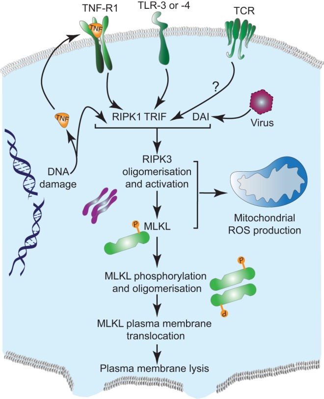
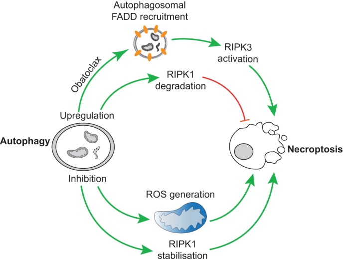
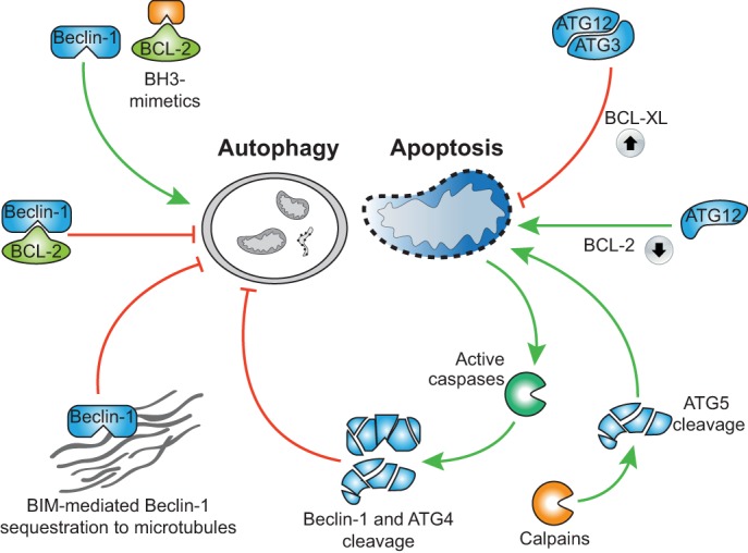
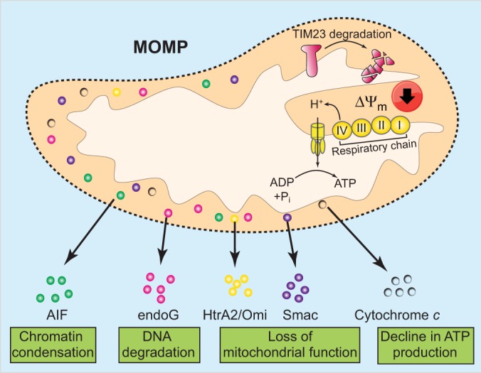
Regulated, programmed cell death is crucial for all multicellular organisms. Cell death is essential in many processes, including tissue sculpting during embryogenesis, development of the immune system and destruction of damaged cells. The best-studied form of programmed cell death is apoptosis, a process that requires activation of caspase proteases. Recently it has been appreciated that various non-apoptotic forms of cell death also exist, such as necroptosis and pyroptosis. These non-apoptotic cell death modalities can be either triggered independently of apoptosis or are engaged should apoptosis fail to execute. In this Commentary, we discuss several regulated non-apoptotic forms of cell death including necroptosis, autophagic cell death, pyroptosis and caspase-independent cell death. We outline what we know about their mechanism, potential roles in vivo and define outstanding questions. Finally, we review data arguing that the means by which a cell dies actually matters, focusing our discussion on inflammatory aspects of cell death.

Keywords: MLKL; Mitochondria; Necroptosis; Pyroptosis; RIPK3.


Figures

Fig. 1.
Fig. 1.
Mechanisms of RIPK3-mediated necroptosis. Various stimuli including DNA damage, viral infection, engagement of receptors, such as TCR, TLR or TNFR, lead to RIPK3 activation. Activation of RIPK3 leads to its oligomerisation and downstream phosphorylation of MLKL. This also results in mitochondria-dependent ROS production. Upon phosphorylation, MLKL oligomerises and translocates to the plasma membrane and causes its lysis.
Fig. 2.
Fig. 2.
Interplay between autophagy and necroptosis. Upregulation of autophagy by Obatoclax can lead to FADD recruitment to autophagosomal membranes and RIPK3 activation resulting in necroptosis (upper part). Conversely, upregulation of autophagy can also lead to RIPK1 degradation and inhibition of necroptosis (lower part). Stimulation of autophagy, but inhibiting its completion, leads to RIPK1 stabilisation and ROS production, both of which contribute to the induction of necroptosis.
Fig. 3.
Fig. 3.
Interplay between autophagy and apoptosis. BCL-2 sequesters Beclin-1, thereby inhibiting autophagy that can be reversed by BH3 mimetics. Furthermore, Bim can sequester Beclin-1 onto microtubules, which also inhibits autophagy (shown on the left). In contrast, ATG12 conjugation to ATG3 enhances BCL-XL expression, thereby inhibiting apoptosis. Unconjugated ATG12 can act in a manner like BH3-only proteins and induce mitochondrial-dependent apoptosis. Calpain-mediated cleavage of ATG5 results in the liberation of a proapoptotic fragment of ATG5 that leads to mitochondrial outer membrane permeabilisation and cell death. In addition, caspase-mediated cleavage of autophagy proteins inhibits autophagy (shown on the right).
Fig. 4.
Fig. 4.
Mechanism of mitochondrial permeabilisation-mediated caspase-independent cell death. Following mitochondrial outer membrane permeabilisation, various intermembrane-space proteins such as Smac, HtrA2/Omi and cytochrome c are released into the cytoplasm. Some of these, such as AIF and endoG, might actively kill the cell in a caspase-independent manner. Over time, mitochondrial functions also decline following outer membrane permeabilisation. This dysfunction includes a loss of mitochondrial protein import due to cleavage of TIM23 and degradation of cytochrome c. Collectively, these events lead to progressive loss of respiratory chain function (complex I to IV) and reduced mitochondrial membrane potential (ΔΨm). Ultimately, this leads to bioenergetic crisis and cell death.

References

    1. Arnoult D., Gaume B., Karbowski M., Sharpe J. C., Cecconi F., Youle R. J. (2003). Mitochondrial release of AIF and EndoG requires caspase activation downstream of Bax/Bak-mediated permeabilization. EMBO J. 22, 4385–4399 10.1093/emboj/cdg423 - DOI - PMC - PubMed
    1. Bahi N., Zhang J., Llovera M., Ballester M., Comella J. X., Sanchis D. (2006). Switch from caspase-dependent to caspase-independent death during heart development: essential role of endonuclease G in ischemia-induced DNA processing of differentiated cardiomyocytes. J. Biol. Chem. 281, 22943–22952 10.1074/jbc.M601025200 - DOI - PubMed
    1. Basit F., Cristofanon S., Fulda S. (2013). Obatoclax (GX15-070) triggers necroptosis by promoting the assembly of the necrosome on autophagosomal membranes. Cell Death Differ. 20, 1161–1173 10.1038/cdd.2013.45 - DOI - PMC - PubMed
    1. Bergsbaken T., Cookson B. T. (2007). Macrophage activation redirects yersinia-infected host cell death from apoptosis to caspase-1-dependent pyroptosis. PLoS Pathog. 3, e161 10.1371/journal.ppat.0030161 - DOI - PMC - PubMed
    1. Bergsbaken T., Fink S. L., Cookson B. T. (2009). Pyroptosis: host cell death and inflammation. Nat. Rev. Microbiol. 7, 99–109 10.1038/nrmicro2070 - DOI - PMC - PubMed

Publication types