. 2014 May 15;55(6):3669-80.
doi: 10.1167/iovs.13-13099.

Mutant ELOVL4 that causes autosomal dominant stargardt-3 macular dystrophy is misrouted to rod outer segment disks

Affiliations

Mutant ELOVL4 that causes autosomal dominant stargardt-3 macular dystrophy is misrouted to rod outer segment disks

Martin-Paul Agbaga et al. Invest Ophthalmol Vis Sci. .

Abstract

Purpose: Autosomal dominant Stargardt macular dystrophy caused by mutations in the Elongation of Very Long Chain fatty acids (ELOVL4) gene results in macular degeneration, leading to early childhood blindness. Transgenic mice and pigs expressing mutant ELOVL4 develop progressive photoreceptor degeneration. The mechanism by which these mutations cause macular degeneration remains unclear, but have been hypothesized to involve the loss of an ER-retention dilysine motif located in the extreme C-terminus. Dominant negative mechanisms and reduction in retinal polyunsaturated fatty acids also have been suggested. To understand the molecular mechanisms involved in disease progression in vivo, we addressed the hypothesis that the disease-linked C-terminal truncation mutant of ELOVL4 exerts a dominant negative effect on wild-type (WT) ELOVL4, altering its subcellular localization and function, which subsequently induces retinal degeneration and loss of vision.

Methods: We generated transgenic Xenopus laevis that overexpress HA-tagged murine ELOVL4 variants in rod photoreceptors.

Results: Tagged or untagged WT ELOVL4 localized primarily to inner segments. However, the mutant protein lacking the dilysine motif was mislocalized to post-Golgi compartments and outer segment disks. Coexpression of mutant and WT ELOVL4 in rods did not result in mislocalization of the WT protein to outer segments or in the formation of aggregates. Full-length HA-tagged ELOVL4 lacking the dilysine motif (K308R/K310R) necessary for targeting the WT ELOVL4 protein to the endoplasmic reticulum was similarly mislocalized to outer segments.

Conclusions: We propose that expression and outer segment mislocalization of the disease-linked 5-base-pair deletion mutant ELOVL4 protein alters photoreceptor structure and function, which subsequently results in retinal degeneration, and suggest three possible mechanisms by which mutant ELOVL4 may induce retinal degeneration in STGD3.

Keywords: autosomal dominant Stargardt-like macular dystrophy (STGD3); elongation of very long chain fatty acids-4 (ELOVL4); photoreceptor outer segment; retinal degeneration.


Figures

Figure 1
Figure 1
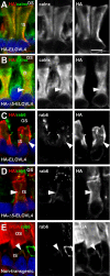
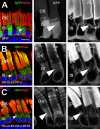
Confocal images of transgenic X. laevis rod photoreceptors expressing WT ELOVL4 (untagged), HA-tagged WT ELOVL4, HA tagged mutant ELOVL4, and rhodopsin-Q344Ter transgenes. (A, B) Merged confocal images of untagged WT-ELOVL4 signal ([A] ELOVL4: green, n = 5) or HA-tagged WT ELOVL4 ([B] HA: green, n = 8), Texas-Red WGA (red) and Hoechst 33342 dye (Nuclei, blue). WT-ELOVL4 and HA-ELOVL4 were identically exclusively localized to rod photoreceptor IS membranes, but excluded from WGA-positive internal membranes of the IS (white arrowhead). WT-ELOVL4 and HA-ELOVL4 were not found within OSs, inner nuclear layer (INL), or ganglion cell layer (GCL). (C) HA-Δ5-ELOVL4 (HA: green, n = 19) was misrouted from rod IS membranes to WGA-positive internal membranes of the IS (white arrowhead) and OS membranes. Colocalization of red and green signals appears yellow/orange. (D) Human rhodopsin-Q344Ter labeled with anti-rhodopsin monoclonal antibody 2B2 (hRho: green, n = 5) was found in rod OS, WGA-positive internal membranes (white arrowhead), lateral plasma membrane (blue arrowhead), and synaptic region plasma membrane (yellow arrowhead). (E) WT X. laevis rhodopsin labeled with monoclonal antibody B630N (xRho: green, n = 3) in X. laevis rod photoreceptors. Arrowheads as in (D). Grayscale images represent nonmerged WGA or antibody signals shown at higher magnification. White arrowheads = WGA-positive internal membranes. Blue arrowheads = plasma membrane. Yellow arrowheads = synaptic region plasma membranes. Scale bar: 50 μm (left), 10 μm (middle), 10 μm (grayscale).
Figure 2
Figure 2
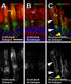
Colocalization of the ER marker calnexin, and Golgi compartment marker rab6, with HA-ELOVL4 and HA-Δ5-ELOVL4 in rod photoreceptor cells. (A) Colocalization of HA-ELOVL4 and ER marker calnexin (HA: red, calnx: green, n = 3) within IS membranes of X. laevis rod photoreceptors. Note complete overlap of calnexin and HA-ELOVL4 signal. (B) Colocalization of HA-Δ5-ELOVL4 and calnexin (n = 3). Note partial overlap of calnexin and HA-Δ5-ELOVL4 signals. Arrowhead (B) indicates region of nonoverlap (HA-positive, calnexin negative). (C) Lack of colocalization of HA-ELOVL4 and Golgi marker, rab6 (HA: red, rab6: green, n = 3) within rod photoreceptor IS membranes. HA-ELOVL4 and rab6 do not colocalize. Arrowhead indicates HA-negative, rab6-positive membranes. (D) Colocalization of HA-Δ5-ELOVL4 and rab6 (HA: red, rab6: green, n = 3). HA-Δ5-ELOVL4 is misrouted to Golgi compartments resulting in significant overlap with rab6 within IS membranes of rod photoreceptor cells (arrowhead). (E) Colocalization of WGA and rab6 labeling (n = 3) in internal membranes of WT rod ISs. The signals are largely coincident within internal membranes of the IS (arrowhead), indicating that most of these membranes are Golgi. Grayscale images show the indicated isolated signals. Scale bars: 5 μm.
Figure 3
Figure 3
HA-Δ5-ELOVL4 is partially delocalized to the OS, but not to plasma membrane and synaptic terminals. (A, B) Confocal micrographs of transgenic X. laevis rod photoreceptors expressing HA-Δ5-ELOVL4 ([A] HA: green, n = 3) labeled in the presence of detergent that reveals delocalization of HA-Δ5-ELOVL4 to OS without prominent plasma membrane labeling. (B) In absence of detergent, there is no observable OS or plasma membrane localization for HA-Δ5-ELOVL4 (HA: green, n = 3). (C) In contrast, in the absence of detergent, 2B2-labeling shows prominent delocalization of Rhodopsin-Q344Ter (hRho: green, n = 3) to OS and lateral plasma membranes (white arrowheads) and synaptic region plasma membrane (yellow arrowhead) of rod photoreceptors. In all images, nuclei were stained with Hoescht 33342 and photoreceptor membranes were labeled with WGA (red). Scale bar: 10 μm.
Figure 4
Figure 4
Effect of coexpression of HA-Δ5-ELOVL4 and WT ELOVL4 on mislocalization of WT ELOVL4 to photoreceptor OS. (AC) Confocal micrographs of X. laevis rod photoreceptor cells coexpressing WT ELOVL4 (ELOVL4, red) and HA-Δ5-ELOVL4 (HA, green) with Hoescht 33342 (blue) (n = 5). WT ELOVL4 expression was restricted to IS without any OS localization (A, C), whereas HA-Δ5-ELOVL4 was distributed within IS and OS membranes (A, B). White arrowheads indicate internal IS membranes (likely Golgi) that are HA-positive and ELOVL4-negative. Left and center are from different transgenic retinas. Right shows higher magnification. Scale bars: 4 and 10 μm.
Figure 5
Figure 5
Role of the ELOVL4 KXKXX motif for inner segment targeting/retention of ELOVL4. (A) Confocal micrographs of transgenic X. laevis rod photoreceptors expressing HA-ELOVL4-KK-RR (HA: green, n = 7); note partial delocalization to OS membranes. Arrowhead indicates WGA-positive IS membranes. (B) Addition of KXKXX motif restores IS localization of HA-Δ5-ELOVL4 in transgenic X. laevis rod photoreceptors expressing HA-Δ5-ELOVL4-ERS (HA: green, n = 8). Grayscale shows the isolated WGA and HA signals. Scale bars: 10 μm.
Figure 6
Figure 6
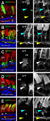
The ELOVL4 KXKXX motif can override OS targeting signals, provided it is located at the extreme carboxyl terminus. (A) Confocal micrograph of rod photoreceptors expressing Rho-GFP-CT (GFP: green, n = 5), which is primarily targeted to rod OS membranes due to presence of the rhodopsin OS targeting motif VXPX. There was no significant IS lateral plasma membrane (blue arrow) or synaptic region plasma membrane (yellow arrow) associated with Rho-GFP-CT. (B) Deletion of the rhodopsin OS targeting motif VXPX caused Rho-GFP-ΔCT signal (GFP: green, n = 5) to be distributed not only to OS, but also to the plasma membrane of the IS, including the lateral plasma membrane (blue arrow) and synaptic region plasma membrane (yellow arrow). (C, D) Addition of the last 12 amino acids containing the ER retention/retrieval signal of ELOVL4 to Rho-GFP-ΔCT and Rho-GFP-CT to generate Rho-GFP-ΔCT-ERS and Rho-GFP-CT-ERS fusion proteins (GFP: green, n = 8) resulted in complete and efficient IS retention and targeting of the fusion proteins. The fusion proteins were excluded from WGA-positive internal membranes (white arrowheads). (E) Rhodopsin OS targeting signal VXPX overrides the KXKXX motif when placed downstream of the KXKXX motif and directs Rho-GFP-CT-ERS-CT fusion protein (GFP: green, n = 5) to photoreceptor OS. No Rho-GFP-CT-ERS-CT fusion protein is retained within IS, including the lateral plasma membrane (blue arrow) or synaptic region membranes (yellow arrow). Scale bars: 10 μm (color) and 5 μm (grayscale).
Figure 7
Figure 7
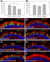
ELOVL4 transgenes are relatively nontoxic, but occasionally induce RD in X. laevis retina. (A, B) Quantification of rod loss in HA-ELOVL4 versus HA-Δ5-ELOVL4 animals using a dot blot assay for rod opsin. There was no statistically significant reduction in rod opsin in X. laevis expressing HA-ELOVL4 versus HA-Δ5-ELOVL4 (A), indicating that neither construct was dramatically more toxic than the other. In contrast, expression of N15S rhodopsin causes a significant decrease in total rod opsin relative to HA-ELOVL4 (P = 0.008, n = 22 per group, Kruskal-Wallace followed by multiple comparisons according to Conover41). (B) Quantification of rod loss in animals expressing GFP, HA-Δ5ELOVL4, and HA-Δ5ELOVL4-3H3Q using dot blot for rod opsin. GFP is a relatively nontoxic transgene product that does not cause retinal degeneration. Total rod opsin levels are not significantly different between groups, indicating that HA-Δ5ELOVL4 and HA-Δ5ELOVL4-3H3Q are not significantly more toxic than GFP, or than each other, although a trend toward RD is apparent for HA-Δ5-ELOVL4 (P = 0.45, n = 27 per group, Kruskal-Wallace). (CJ) Expression of transgenic GFP (GFP: green, n = 7) (C) did not cause retinal degeneration and retained photoreceptor integrity similar to nontransgenics (J). However, occasionally retinas expressing ELOVL4 variants (DH) had RD apparent by histology, as indicated by both loss of rods and shortened rod outer segments (n as reported in Figs. 1, 5, 8). Scale bar: 50 μm.
Figure 8
Figure 8
Localization of histidine-deficient mutant ELOVL4. (A) Transgenic expressed GFP protein (GFP: green, n = 7) is present throughout the IS, and at lower concentrations in OS. (B) Transgenically expressed HA-Δ5-ELOVL4 (HA: green, n = 19) and (C) HA-Δ5-ELOVL4-3H3Q HA: green, n = 10) fusion proteins were both localized to IS membranes, including WGA-positive internal membranes (arrowheads), and to photoreceptor OSs. Scale bars: 10 μm (merged) or 5 μm (grayscale).

Similar articles

Cited by

References

    1. Zhang K, Kniazeva M, Han M, et al. A 5-bp deletion in ELOVL4 is associated with two related forms of autosomal dominant macular dystrophy. Nat Genet. 2001; 27: 89–93 - PubMed
    1. Agbaga MP, Brush RS, Mandal MN, Elliott MH, Al-Ubaidi MR, Anderson RE. Role of elovl4 protein in the biosynthesis of docosahexaenoic acid. Adv Exp Med Biol. 2010; 664: 233–242 - PMC - PubMed
    1. Carmona-Antonanzas G, Monroig O, Dick JR, Davie A, Tocher DR. Biosynthesis of very long-chain fatty acids (C>24) in Atlantic salmon: cloning, functional characterisation, and tissue distribution of an Elovl4 elongase. Comp Biochem Physiol B Biochem Mol Biol. 2011; 159: 122–129 - PubMed
    1. Ambasudhan R, Wang X, Jablonski MM, et al. Atrophic macular degeneration mutations in ELOVL4 result in the intracellular misrouting of the protein. Genomics. 2004; 83: 615–625 - PubMed
    1. Karan G, Yang Z, Zhang K. Expression of wild type and mutant ELOVL4 in cell culture: subcellular localization and cell viability. Mol Vis. 2004; 10: 248–253 - PubMed

Publication types

MeSH terms

Supplementary concepts