. 2014 May 6:8:26.
doi: 10.3389/fnana.2014.00026. eCollection 2014.

Investigation of spinal cerebrospinal fluid-contacting neurons expressing PKD2L1: evidence for a conserved system from fish to primates

Affiliations

Investigation of spinal cerebrospinal fluid-contacting neurons expressing PKD2L1: evidence for a conserved system from fish to primates

Lydia Djenoune et al. Front Neuroanat. .

Abstract

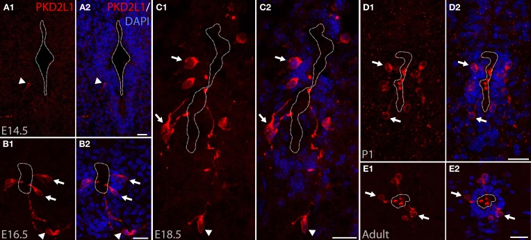
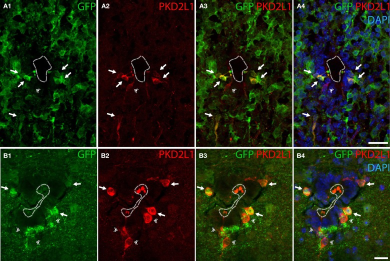
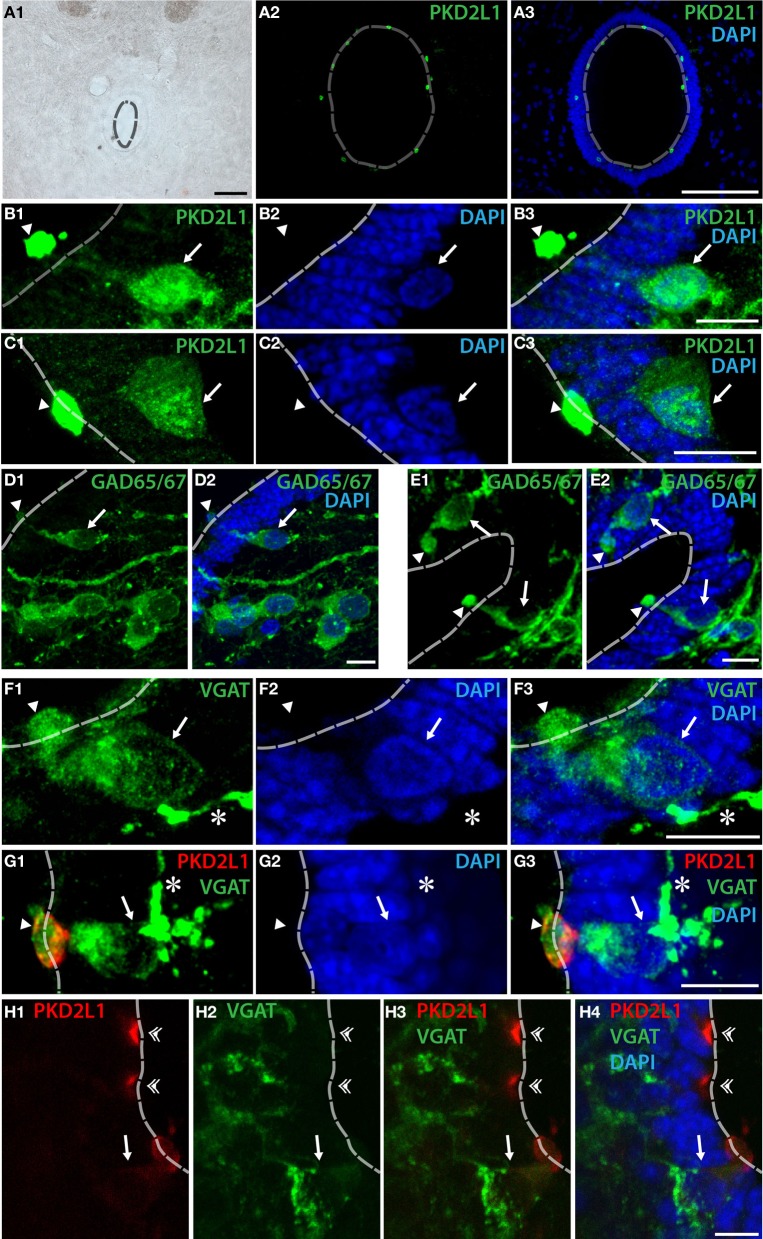
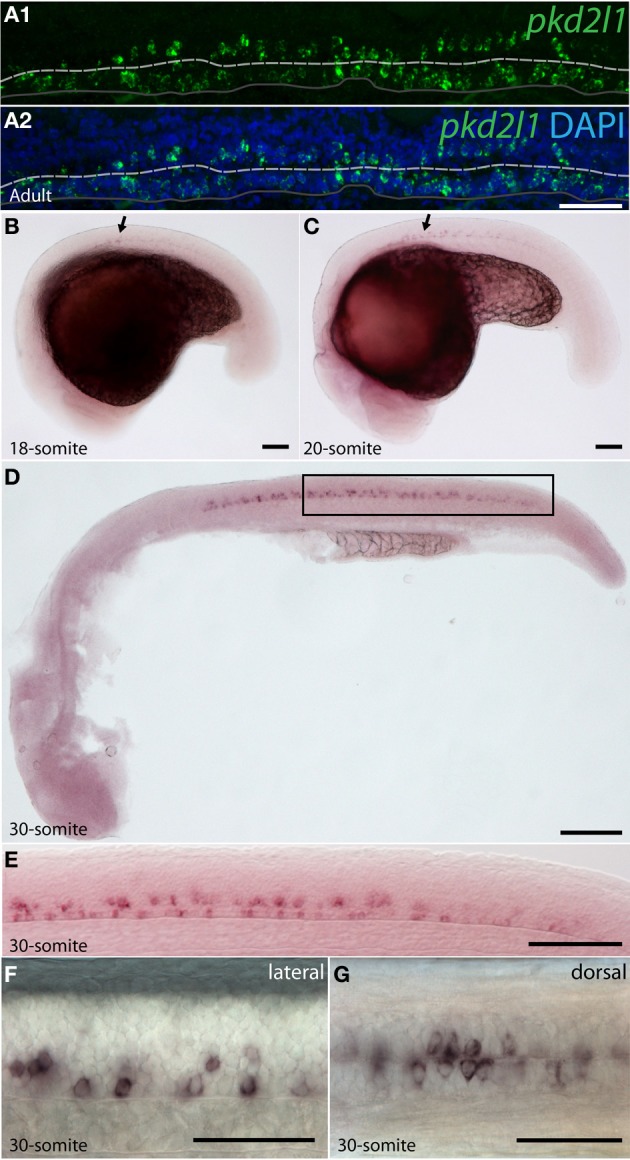
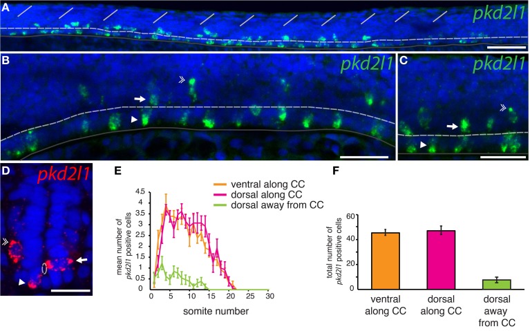
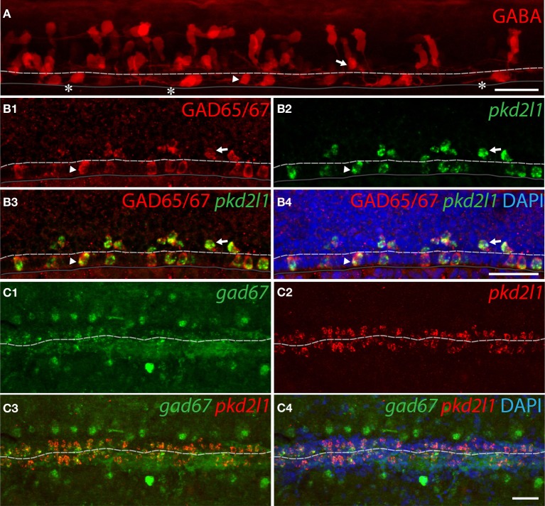
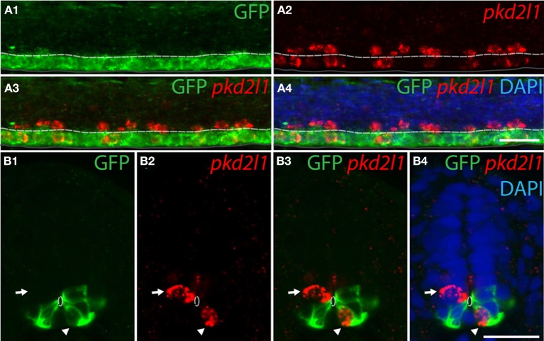
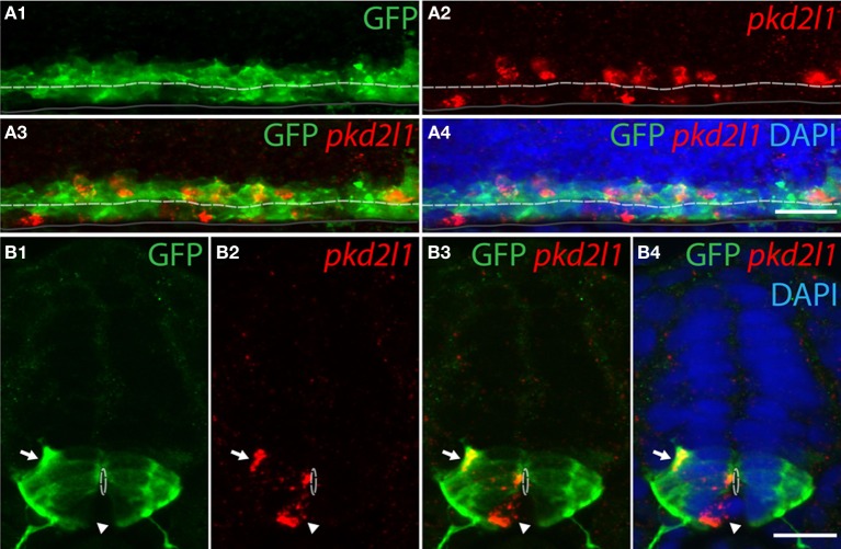
Over 90 years ago, Kolmer and Agduhr identified spinal cerebrospinal fluid-contacting neurons (CSF-cNs) based on their morphology and location within the spinal cord. In more than 200 vertebrate species, they observed ciliated neurons around the central canal that extended a brush of microvilli into the cerebrospinal fluid (CSF). Although their morphology is suggestive of a primitive sensory cell, their function within the vertebrate spinal cord remains unknown. The identification of specific molecular markers for these neurons in vertebrates would benefit the investigation of their physiological roles. PKD2L1, a transient receptor potential channel that could play a role as a sensory receptor, has been found in cells contacting the central canal in mouse. In this study, we demonstrate that PKD2L1 is a specific marker for CSF-cNs in the spinal cord of mouse (Mus musculus), macaque (Macaca fascicularis) and zebrafish (Danio rerio). In these species, the somata of spinal PKD2L1(+) CSF-cNs were located below or within the ependymal layer and extended an apical bulbous extension into the central canal. We found GABAergic PKD2L1-expressing CSF-cNs in all three species. We took advantage of the zebrafish embryo for its transparency and rapid development to identify the progenitor domains from which pkd2l1 (+) CSF-cNs originate. pkd2l1 (+) CSF-cNs were all GABAergic and organized in two rows-one ventral and one dorsal to the central canal. Their location and marker expression is consistent with previously described Kolmer-Agduhr cells. Accordingly, pkd2l1 (+) CSF-cNs were derived from the progenitor domains p3 and pMN defined by the expression of nkx2.2a and olig2 transcription factors, respectively. Altogether our results suggest that a system of CSF-cNs expressing the PKD2L1 channel is conserved in the spinal cord across bony vertebrate species.

Keywords: GABAergic neurons; PKD2L1; cerebrospinal fluid-contacting neurons (CSF-cNs); macaque; mouse; spinal cord; zebrafish.


Figures

Figure 1
Figure 1
PKD2L1 expression in CSF-cNs is present from embryonic stage E14.5 to adulthood in mouse. (A–E) Immunostaining for PKD2L1 at E14.5 (A), E16.5 (B), E18.5 (C), P1 (D), and in the adult (E) performed on coronal sections of the mouse cervical spinal cord. At E14.5, few PKD2L1+ cells are detected (arrowhead). From E16.5 (B) to adult (E), multiple PKD2L1+ CSF-cNs (arrows) surround the central canal where they project an apical bulbous dendritic extension contacting its lumen. Note that PKD2L1+ cells exhibiting a similar morphology but not distinctly contacting the central canal (arrowheads) can also be observed (A–C). Dorsal is up. White dashed line delineates the central canal. DAPI staining appears in blue. Scale bars = 20 μm.
Figure 2
Figure 2
Some PKD2L1+ cells surrounding the central canal of cervical spinal cord of adult mouse are immunoreactive for GFP in the knock-in GAD67-GFP mouse. (A) 10 μm-thick coronal sections of GAD67-GFP E16.5 mouse spinal cord immunostained for GFP (A1,A3,A4) and PKD2L1 (A2–A4). Most PKD2L1+ cells are GFP+ (arrows) but not all (double arrowheads). (B) 30 μm-thick coronal sections of a GAD67-GFP adult mouse spinal cord immunostained for GFP (B1,B3,B4) and PKD2L1 (B2–B4). While many PKD2L1+cells are distinctly GFP+ (arrows), some cells are not (double arrowheads). Dorsal is up. White dashed line delineates the central canal. DAPI staining appears in blue. Scale bars = 25 μm (A), 15 μm (B).
Figure 3
Figure 3
PKD2L1+ CSF-cNs can be found as GABAergic neurons surrounding the central canal in the spinal cord of the adult macaque. (A–C) PKD2L1 immunostaining labels intraluminal buds (A–C) and rarely subependymal or ependymal cell bodies (B,C). (D,E) GAD65/67 immunoreactive cells are numerous in the vicinity of the central canal: proximal cells show a clear apical extension contacting the lumen while cells located away do not. (F) CSF-cNs surrounding the central canal are immunoreactive for the vesicular GABA transporter VGAT. (G,H) Double immunostaining for VGAT and PKD2L1 shows that some but not all PKD2L1+ CSF-cNs are clearly GABAergic. The intraluminal buds can be found double-labeled for PKD2L1 and VGAT (arrowheads) but not systematically (double arrowheads), while apical extensions, cell body and putative axon are usually only labeled by VGAT. All panels are coronal sections of the macaque adult spinal cord taken at the lumbar level for (A,G), at the cervical level for (B–F) and at the thoracic level for (H). In all panels, arrows point to the somata, asterisks to the putative axons and arrowheads to the intraluminal buds of CSF-cNs. White dashed line delineates the central canal. DAPI staining appears in blue. Scale bars = 100 μm (A) and 10 μm (B–H).
Figure 4
Figure 4
pkd2l1 is expressed from the embryonic 18-somite stage to adulthood in the zebrafish spinal cord. (A) Adult; (B–G) embryonic stages: 18-somite (B), 20-somite (C) and 30-somite stages (D–G). (A) In sagittal sections from the adult spinal cord, pkd2l1+ CSF-contacting cells are located ventral and dorsal to the central canal. White dash line: central canal, dark gray line: ventral limit of the spinal cord. pkd2l1 expression appears in the rostral spinal cord (B,C, arrows) and is distributed in two rows of cells along the rostro-caudal axis (E,F) and on each side of the midline (G). (E) is a close-up of the black box from (D). Rostral is to the left for all panels. Lateral views with dorsal up for (A–F) and dorsal view for (G). DAPI staining appears in blue. Scale bars = 40 μm (A), 100 μm for (B–D), 50 μm for (E–G).
Figure 5
Figure 5
pkd2l1 is expressed in cells contacting the CSF and surrounding the central canal in the spinal cord of the zebrafish embryo. (A–D) FISH of pkd2l1 mRNA at the 30-somite stage. Lateral views for (A–C) and transverse section for (D). Two subpopulations of pkd2l1 bright expressing cells surround the central canal ventrally (arrowhead) and dorsally (arrow). Note the existence of dorsal pkd2l1 weak expressing cells away from the central canal (double arrowhead). The white dashed line, dark gray solid line and the solid white lines indicate respectively the central canal, the ventral limit of the spinal cord, and the somite boundaries. DAPI staining appears in blue. Scale bars = 50 μm for (A), 30 μm for (B,C), and 20 μm for (D). (E) Mean number of pkd2l1+ cells per somite along the rostro-caudal axis at the 30-somite stage. (F) Total cell counts per embryo (n = 13 embryos). We counted 45.4 ± 2.4 ventral pkd2l1+ cells, 47.1 ± 3.4 dorsal pkd2l1+ cells and 7.6 ± 2.3 dorsal away from the central canal pkd2l1+ cells per embryo. (E,F) Mean values are given ± s.e.m.
Figure 6
Figure 6
pkd2l1+ CSF-cNs are GABAergic neurons expressing GABA and GAD in zebrafish. (A) GABA IHC on WT 30-somite embryos shows ventral (arrowhead) and dorsal (arrow) KAs and their ascending axon (asterisks). (B1–B4) Ventral and dorsal pkd2l1+ KAs are GAD65/67 immunoreactive in 30-somite embryos as shown by FISH for pkd2l1 (green) coupled to a GAD65/67 IHC (red). (C1–C4) In the adult, pkd2l1+ KAs are gad67+ as shown by FISH for gad67 (C1,C3,C4) and pkd2l1 (C2–C4) on sections of WT spinal cord. The white dash line indicates the central canal, the dark line the ventral limit of the spinal cord. (A) is a projection from the lateral view of a whole-mount embryo immunostained for GABA while (B1–B4) correspond to sagittal sections and (C1–C4) to frontal sections. DAPI staining appears in blue. Scale bars = 30 μm.
Figure 7
Figure 7
Ventral pkd2l1+ cells express nkx2.2a and derive from the p3 progenitor domain in the zebrafish embryo. (A,B) pkd2l1 FISH in Tg(nkx2.2a: mEGFP) embryos immunostained for GFP at the 30-somite stage. IHC for GFP reveals the p3 domain. (A) Lateral view showing that the most ventral pkd2l1+ cells are GFP+. (B) A typical transverse section shows a ventral pkd2l1+ cell contacting the central canal and expressing GFP (arrowhead) while a dorsal pkd2l1+ cell contacting the central canal does not express GFP (arrow) in the Tg(nkx2.2a: mEGFP) transgenic embryo. White dashed line delineates the central canal, dark line the ventral limit of the spinal cord. DAPI staining appears in blue. Scale bars = 20 μm.
Figure 8
Figure 8
Dorsal pkd2l1+ CSF-cNs express olig2 and derive from the pMN progenitor domain in the embryonic spinal cord of zebrafish. (A,B) pkd2l1 FISH in Tg(olig2:EGFP) embryos immunostained for GFP at the 30-somite stage. IHC for GFP reveals the pMN domain. (A) Lateral view showing dorsal pkd2l1+ cells express GFP. (B) Transverse sections showing dorsal pkd2l1+ cells contacting the central canal express GFP (arrow) while a ventral pkd2l1+ cell contacting the central canal (arrowhead) does not express GFP in the Tg(olig2:EGFP) transgenic line. White dashed line delineates the central canal, dark line the ventral limit of the spinal cord. DAPI staining appears in blue. Scale bars = 20 μm.

References

    1. Acerbo M. J., Hellmann B., Gunturkun O. (2003). Catecholaminergic and dopamine-containing neurons in the spinal cord of pigeons: an immunohistochemical study. J. Chem. Neuroanat. 25, 19–27 10.1016/S0891-0618(02)00072-8 - DOI - PubMed
    1. Agduhr E. (1922). Über ein Zentrales Sinnesorgan (?) bei den Vertebraten. Z. Anat. Entwicklungs. 66, 223–360 10.1007/BF02593586 - DOI
    1. Alunni A., Krecsmarik M., Bosco A., Galant S., Pan L., Moens C. B., et al. (2013). Notch3 signaling gates cell cycle entry and limits neural stem cell amplification in the adult pallium. Development 140, 3335–3347 10.1242/dev.095018 - DOI - PMC - PubMed
    1. Barber R. P., Vaughn J. E., Roberts E. (1982). The cytoarchitecture of GABAergic neurons in rat spinal cord. Brain Res. 238, 305–328 10.1016/0006-8993(82)90107-X - DOI - PubMed
    1. Basora N., Nomura H., Berger U. V., Stayner C., Guo L., Shen X., et al. (2002). Tissue and cellular localization of a novel polycystic kidney disease-like gene product, polycystin-L. J. Am. Soc. Nephrol. 13, 293–301 - PubMed

LinkOut - more resources