

Myocardial T1 mapping: modalities and clinical applications
- PMID: 24834410
- PMCID: PMC3996234
- DOI: 10.3978/j.issn.2223-3652.2013.09.03
Myocardial T1 mapping: modalities and clinical applications
Abstract
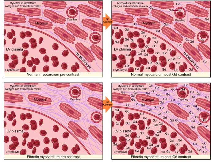
Myocardial fibrosis appears to be linked to myocardial dysfunction in a multitude of non-ischemic cardiomyopathies. Accurate non-invasive quantitation of this extra-cellular matrix has the potential for widespread clinical benefit in both diagnosis and guiding therapeutic intervention. T1 mapping is a cardiac magnetic resonance (CMR) imaging technique, which shows early clinical promise particularly in the setting of diffuse fibrosis. This review will outline the evolution of T1 mapping and the various techniques available with their inherent advantages and limitations. Histological validation of this technique remains somewhat limited, however clinical application in a range of pathologies suggests strong potential for future development.
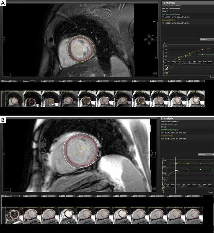
Keywords: T1 mapping; extracellular volume; modified Look Locker inversion recovery (MOLLI); myocardial fibrosis.
Figures
References
-
- Sugihara N, Genda A, Shimizu M, et al. Diastolic dysfunction and its relation to myocardial fibrosis in essential hypertension. J Cardiol 1988;18:353-61 - PubMed
-
- Ling LH, Kistler PM, Ellims AH, et al. Diffuse ventricular fibrosis in atrial fibrillation: noninvasive evaluation and relationships with aging and systolic dysfunction. J Am Coll Cardiol 2012;60:2402-8 - PubMed
-
- McLenachan JM, Dargie HJ. Ventricular arrhythmias in hypertensive left ventricular hypertrophy. Relationship to coronary artery disease, left ventricular dysfunction, and myocardial fibrosis. Am J Hypertens 1990;3:735-40 - PubMed
-
- Picano E, Pelosi G, Marzilli M, et al. In vivo quantitative ultrasonic evaluation of myocardial fibrosis in humans. Circulation 1990;81:58-64 - PubMed
-
- Knaapen P, Boellaard R, Götte MJ, et al. Perfusable tissue index as a potential marker of fibrosis in patients with idiopathic dilated cardiomyopathy. J Nucl Med 2004;45:1299-304 - PubMed
Publication types
LinkOut - more resources
Full Text Sources
Other Literature Sources