

Assessing the end-organ in peripheral arterial occlusive disease-from contrast-enhanced ultrasound to blood-oxygen-level-dependent MR imaging
- PMID: 24834413
- PMCID: PMC3996233
- DOI: 10.3978/j.issn.2223-3652.2014.03.02
Assessing the end-organ in peripheral arterial occlusive disease-from contrast-enhanced ultrasound to blood-oxygen-level-dependent MR imaging
Abstract
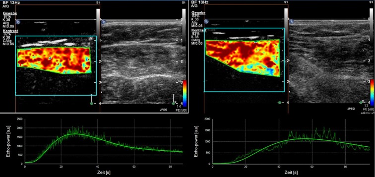
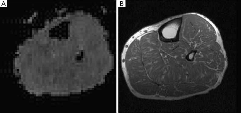
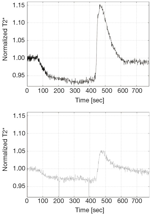
Peripheral arterial occlusive disease (PAOD) is a result of atherosclerotic disease which is currently the leading cause of morbidity and mortality in the western world. Patients with PAOD may present with intermittent claudication or symptoms related to critical limb ischemia. PAOD is associated with increased mortality rates. Stenoses and occlusions are usually detected by macrovascular imaging, including ultrasound and cross-sectional methods. From a pathophysiological view these stenoses and occlusions are affecting the microperfusion in the functional end-organs, such as the skin and skeletal muscle. In the clinical arena new imaging technologies enable the evaluation of the microvasculature. Two technologies currently under investigation for this purpose on the end-organ level in PAOD patients are contrast-enhanced ultrasound (CEUS) and blood-oxygen-level-dependent (BOLD) MR imaging (MRI). The following article is providing an overview about these evolving techniques with a specific focus on skeletal muscle microvasculature imaging in PAOD patients.
Keywords: Duplex ultrasonography; magnetic resonance imaging; microbubbles; peripheral arterial disease.
Figures
Similar articles
-
Blood oxygenation level-dependent magnetic resonance imaging of the skeletal muscle in patients with peripheral arterial occlusive disease.Circulation. 2006 Jun 27;113(25):2929-35. doi: 10.1161/CIRCULATIONAHA.105.605717. Epub 2006 Jun 19. Circulation. 2006. PMID: 16785340
-
Blood oxygenation level-dependent MRI of the skeletal muscle during ischemia in patients with peripheral arterial occlusive disease.Rofo. 2009 Dec;181(12):1157-61. doi: 10.1055/s-0028-1109786. Epub 2009 Oct 26. Rofo. 2009. PMID: 19859866
-
A Reperfusion BOLD-MRI Tissue Perfusion Protocol Reliably Differentiate Patients with Peripheral Arterial Occlusive Disease from Healthy Controls.J Clin Med. 2021 Aug 18;10(16):3643. doi: 10.3390/jcm10163643. J Clin Med. 2021. PMID: 34441939 Free PMC article.
-
Clinical implications of skeletal muscle blood-oxygenation-level-dependent (BOLD) MRI.MAGMA. 2012 Aug;25(4):251-61. doi: 10.1007/s10334-012-0306-y. Epub 2012 Feb 29. MAGMA. 2012. PMID: 22374263 Review.
-
Dynamic contrast-enhanced magnetic resonance imaging: fundamentals and application to the evaluation of the peripheral perfusion.Cardiovasc Diagn Ther. 2014 Apr;4(2):147-64. doi: 10.3978/j.issn.2223-3652.2014.03.01. Cardiovasc Diagn Ther. 2014. PMID: 24834412 Free PMC article. Review.
Cited by
-
Contrast-enhanced ultrasound after endovascular aortic repair-current status and future perspectives.Cardiovasc Diagn Ther. 2015 Dec;5(6):454-63. doi: 10.3978/j.issn.2223-3652.2015.09.04. Cardiovasc Diagn Ther. 2015. PMID: 26673398 Free PMC article. Review.
-
Risk of Peripheral Arterial Occlusive Disease with Periodontitis and Dental Scaling: A Nationwide Population-Based Cohort Study.Int J Environ Res Public Health. 2022 Aug 15;19(16):10057. doi: 10.3390/ijerph191610057. Int J Environ Res Public Health. 2022. PMID: 36011700 Free PMC article.
-
Muscle oxygenation during dynamic plantar flexion exercise: combining BOLD MRI with traditional physiological measurements.Physiol Rep. 2016 Oct;4(20):e13004. doi: 10.14814/phy2.13004. Epub 2016 Oct 24. Physiol Rep. 2016. PMID: 27798357 Free PMC article.
-
Association between psoriasis and peripheral artery occlusive disease: a population-based retrospective cohort study.Front Cardiovasc Med. 2023 Jun 12;10:1136540. doi: 10.3389/fcvm.2023.1136540. eCollection 2023. Front Cardiovasc Med. 2023. PMID: 37378400 Free PMC article.
-
Characterizing blood oxygen level-dependent (BOLD) response following in-magnet quadriceps exercise.MAGMA. 2015 Jun;28(3):271-8. doi: 10.1007/s10334-014-0461-4. Epub 2014 Sep 24. MAGMA. 2015. PMID: 25248947
References
-
- Kröger K, Stang A, Kondratieva J, et al. Prevalence of peripheral arterial disease - results of the Heinz Nixdorf recall study. Eur J Epidemiol 2006;21:279-85 - PubMed
-
- Hirsch AT, Haskal ZJ, Hertzer NR, et al. ACC/AHA 2005 Practice Guidelines for the management of patients with peripheral arterial disease (lower extremity, renal, mesenteric, and abdominal aortic): a collaborative report from the American Association for Vascular Surgery/Society for Vascular Surgery, Society for Cardiovascular Angiography and Interventions, Society for Vascular Medicine and Biology, Society of Interventional Radiology, and the ACC/AHA Task Force on Practice Guidelines (Writing Committee to Develop Guidelines for the Management of Patients With Peripheral Arterial Disease): endorsed by the American Association of Cardiovascular and Pulmonary Rehabilitation; National Heart, Lung, and Blood Institute; Society for Vascular Nursing; TransAtlantic Inter-Society Consensus; and Vascular Disease Foundation. Circulation 2006;113:e463-654 - PubMed
-
- Askew CD, Green S, Walker PJ, et al. Skeletal muscle phenotype is associated with exercise tolerance in patients with peripheral arterial disease. J Vasc Surg 2005;41:802-7 - PubMed
-
- Mitchell RG, Duscha BD, Robbins JL, et al. Increased levels of apoptosis in gastrocnemius skeletal muscle in patients with peripheral arterial disease. Vasc Med 2007;12:285-90 - PubMed
-
- Staub D, Partovi S, Imfeld S, et al. Novel applications of contrast-enhanced ultrasound imaging in vascular medicine. Vasa 2013;42:17-31 - PubMed
Publication types
LinkOut - more resources
Full Text Sources