

A comparative study of thin-section CT findings between seasonal influenza virus pneumonia and Streptococcus pneumoniae pneumonia
- PMID: 24834476
- PMCID: PMC4075588
- DOI: 10.1259/bjr.20140051
A comparative study of thin-section CT findings between seasonal influenza virus pneumonia and Streptococcus pneumoniae pneumonia
Abstract
Objective: To compare the pulmonary thin-section CT findings in patients with seasonal influenza virus pneumonia with Streptococcus pneumoniae pneumonia.
Methods: The study group included 30 patients (20 males and 10 females; age range, 20-91 years; mean age, 55.9 years) with seasonal influenza virus pneumonia and 71 patients (47 males and 24 females; age range, 27-92 years; mean age, 67.5 years) with S. pneumoniae pneumonia.
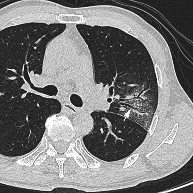
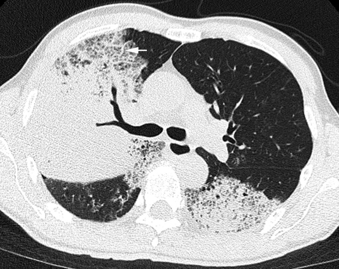
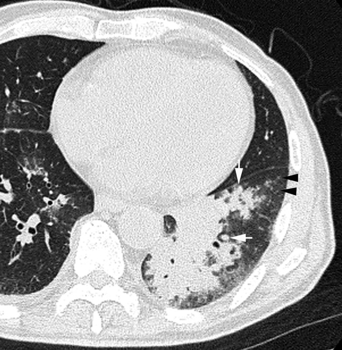
Results: The proportion of community-acquired infection was significantly higher in patients with influenza virus pneumonia than with S. pneumoniae pneumonia (p = 0.001). CT findings of ground-glass attenuation (GGA) (p = 0.012) and crazy-paving appearance (p = 0.03) were significantly more frequent in patients with influenza virus pneumonia than with S. pneumoniae pneumonia. Conversely, consolidation (p < 0.001), mucoid impaction (p < 0.001), centrilobular nodules (p = 0.04) and pleural effusion (p = 0.003) were significantly more frequent in patients with S. pneumoniae pneumonia than in those with influenza virus pneumonia.
Conclusion: Pulmonary thin-section CT findings, such as consolidation and mucoid impaction may be useful in distinguishing between seasonal influenza virus pneumonia and S. pneumoniae pneumonia.
Advances in knowledge: (1) Distinguishing seasonal influenza virus pneumonia with S. pneumoniae pneumonia is important. (2) The CT findings of GGA and crazy-paving appearance were more frequently found in patients with influenza virus pneumonia than in patients with S. pneumoniae pneumonia, whereas consolidation, mucoid impaction, centrilobular nodules and pleural effusion were more frequently found in patients with S. pneumoniae pneumonia.
Figures


References
-
- Craven DE, Steger KA. Epidemiology of nosocomial pneumonia. New perspective on an old disease. Chest 1995; 108(2 Suppl): 1S–16S. - PubMed
-
- Chastre J, Fagon JF. Pneumonia in the ventilator-dependent patient. In: Tobin M, ed. Principles and practice of mechanical ventilation. 1st edn. New York, NY: McGraw & Hill; 1994. pp. 857–90.
-
- American Thoracic Society. Hospital-acquired pneumonia in adults: diagnosis, assessment of severity, initial antimicrobial therapy, and preventive strategies: a consensus statement. Am J Respir Crit Care Med 1996; 153: 1711–25. - PubMed
-
- Dominguez J, Gali N, Blanco S, Pedroso P, Prat C, Matas L, et al. Detection of Streptococcus pneumoniae antigen by a rapid immunochromatographic assay in urine samples. Chest 2001; 119: 243–9. - PubMed
Publication types
MeSH terms
LinkOut - more resources
Full Text Sources
Other Literature Sources
Medical
