Review
. 2014 Sep-Oct;20(5):379-89.
doi: 10.5152/dir.2014.13396.

Perfusion CT imaging of the liver: review of clinical applications

Affiliations
Review

Perfusion CT imaging of the liver: review of clinical applications

Hayri Oğul et al. Diagn Interv Radiol. 2014 Sep-Oct.

Abstract

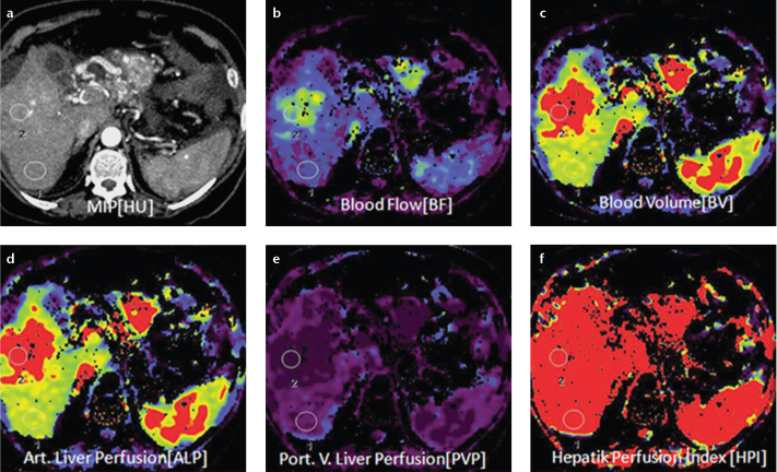
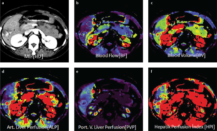
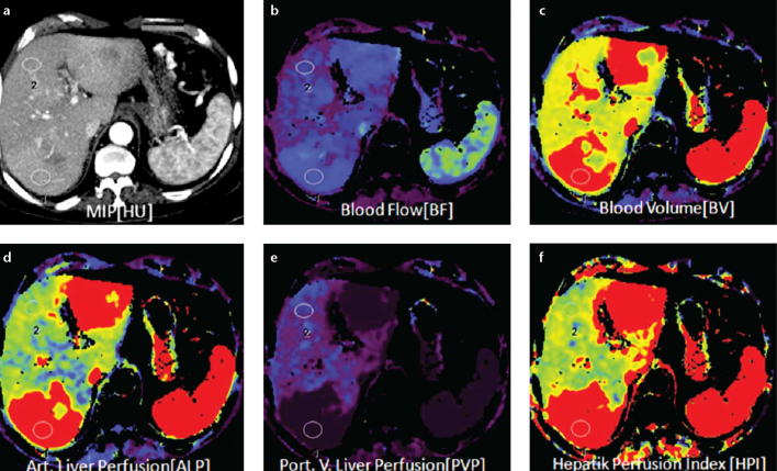
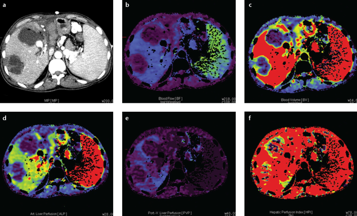
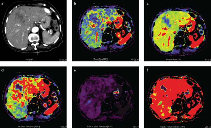
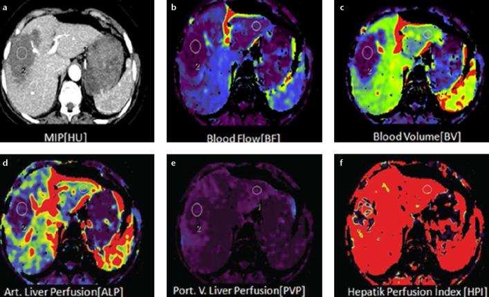
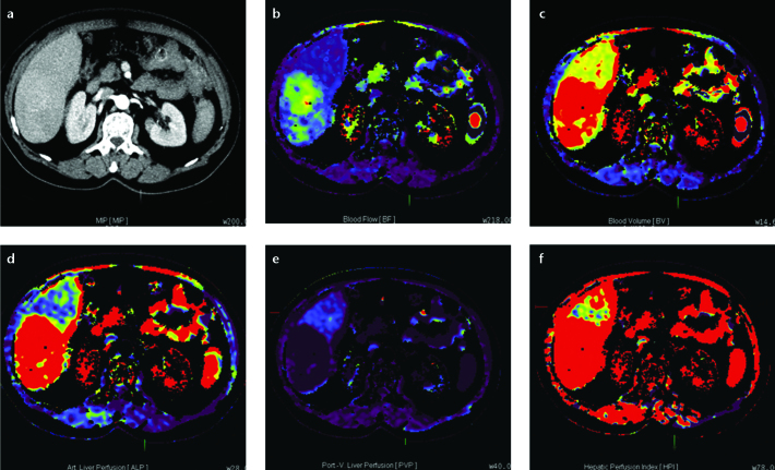
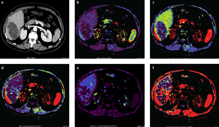
Perfusion computed tomography (CT) has a great potential for determining hepatic and portal blood flow; it offers the advantages of quantitative determination of lesion hemodynamics, distinguishing malignant and benign processes, as well as providing morphological data. Many studies have reported the use of this method in the assessment of hepatic tumors, hepatic fibrosis associated with chronic liver disease, treatment response following radiotherapy and chemotherapy, and hepatic perfusion changes after radiological or surgical interventions. The main goal of liver perfusion imaging is to improve the accuracy in the characterization of liver disorders. In this study, we reviewed the clinical application of perfusion CT in various hepatic diseases.


Figures

Figure 1. a–f.
Figure 1. a–f.
Axial perfusion images (a) and functional CT perfusion map (b–f) of a 60-year-old man with hepatocellular carcinoma in the right lobe of the liver. The tumor manifests as a distinct range of colors compared with the background liver parenchyma. The functional map shows the wash out in the tumor. ALP, arterial liver perfusion; BF, blood flow; BV, blood volume; HPI, hepatic perfusion index; MIP, maximum intensity projection; PLP, portal liver perfusion.
Figure 2. a–f.
Figure 2. a–f.
Axial perfusion images (a) and functional CT perfusion map (b–f) of a 29-year-old woman with hemangioma. The perfusion images show increased BF, ALP and HPI but decreased PLP and BV compared with normal liver parenchyma.
Figure 3. a–f.
Figure 3. a–f.
Axial perfusion images (a) and functional CT perfusion map (b–f) of a 70-year-old woman with liver metastasis from pancreatic adenocarcinoma. The perfusion images show increased BF, BV, and ALP compared with normal liver parenchyma.
Figure 4. a–f.
Figure 4. a–f.
Axial perfusion images (a) and functional CT perfusion map (b–f) of a 39-year-old man with hepatic arterial occlusion after living donor liver transplantation. The perfusion images show decreased BF, BV, ALP, PVP, and HPI at the necrotic area.
Figure 5. a–f.
Figure 5. a–f.
Axial perfusion images (a) and functional CT perfusion map (b–f) of a 45-year-old man with cholangiocarcinoma. The tumor shows an increased hepatic arterial perfusion and decreased portal perfusion compared with the normal parenchyma.
Figure 6. a–f.
Figure 6. a–f.
Axial perfusion images (a) and functional CT perfusion map (b–f) of a 29-year-old man with alveolar echinococcosis. The lesion shows a decreased hepatic arterial perfusion compared with the normal parenchyma.
Figure 7. a–f.
Figure 7. a–f.
Axial perfusion images (a) and functional CT perfusion map (b–f) of a 53-year-old man with hepatocellular carcinoma before chemoembolization. The tumor shows an increased hepatic arterial perfusion and decreased hepatic portal perfusion compared with the normal parenchyma.
Figure 8. a–f.
Figure 8. a–f.
Axial perfusion images (a) and functional CT perfusion map (b–f) of a 53-year-old man (same patient with Fig. 7) with hepatocellular carcinoma after chemoembolization. The tumor shows a decreased hepatic arterial and portal perfusion compared with the normal parenchyma.

References

    1. Kapoor V, Brancatelli G, Federle MP, et al. Multidetector CT arteriography with volumetric three-dimensional rendering to evaluate patients with metastatic colorectal disease for placement of a floxuridine infusion pump. AJR Am J Roentgenol. 2003;181:455–463. - PubMed
    1. Kanda T, Yoshikawa T, Ohno Y, et al. Perfusion measurement of the whole upper abdomen of patients with and without liver diseases: initial experience with 320-detector row CT. Eur J Radiol. 2012;81:2470–2475. - PubMed
    1. Cao X, Jiang X. Evaluating the effect of high-intensity focused ultrasound therapy on liver tumors using multislice CT perfusion. Oncol Lett. 2013;5:511–514. - PMC - PubMed
    1. Kambadakone AR, Sahani DV. Body perfusion CT: technique, clinical applications, and advances. Radiol Clin North Am. 2009;47:161–178. - PubMed
    1. Wang X, Xue HD, Jin ZY, et al. Quantitative hepatic CT perfusion measurement: comparison of Couinaud’s hepatic segments with dual-source 128-slice CT. Eur J Radiol. 2013;82:220–226. - PubMed

MeSH terms