

Lubricin is Required for the Structural Integrity and Post-natal Maintenance of TMJ
- PMID: 24834922
- PMCID: PMC4107552
- DOI: 10.1177/0022034514535807
Lubricin is Required for the Structural Integrity and Post-natal Maintenance of TMJ
Abstract
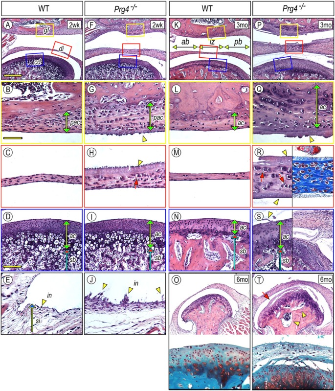
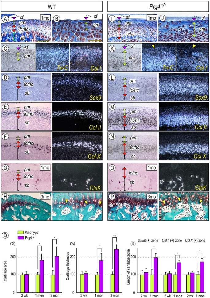
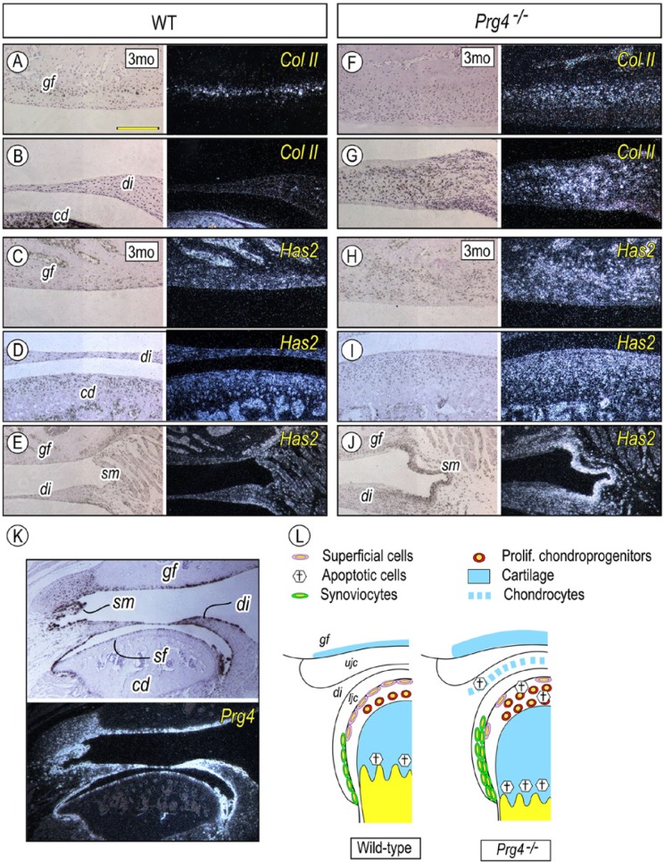
The Proteoglycan 4 (Prg4) product lubricin plays essential roles in boundary lubrication and movement in limb synovial joints, but its roles in temporomandibular joint (TMJ) are unclear. Thus, we characterized the TMJ phenotype in wild-type and Prg4(-/-) mouse littermates over age. As early as 2 weeks of age, mutant mice exhibited hyperplasia in the glenoid fossa articular cartilage, articular disc, and synovial membrane. By 1 month of age, there were fewer condylar superficial tenascin-C/Col1-positive cells and more numerous apoptotic condylar apical cells, while chondroprogenitors displayed higher mitotic activity, and Sox9-, Col2-, and ColX-expressing chondrocyte zones were significantly expanded. Mutant subchondral bone contained numerous Catepsin K-expressing osteoclasts at the chondro-osseous junction, increased invasive marrow cavities, and suboptimal subchondral bone. Mutant glenoid fossa, disc, synovial cells, and condyles displayed higher Hyaluronan synthase 2 expression. Mutant discs also lost their characteristic concave shape, exhibited ectopic chondrocyte differentiation, and occasionally adhered to condylar surfaces. A fibrinoid substance of unclear origin often covered the condylar surface. By 6 months of age, mutant condyles displayed osteoarthritic degradation with apical/mid-zone separation. In sum, lubricin exerts multiple essential direct and indirect roles to preserve TMJ structural and cellular integrity over post-natal life.
Keywords: Has2; Prg4; hyaluronic acid; osteoarthritis; superficial layer; temporomandibular joint.
© International & American Associations for Dental Research.
Conflict of interest statement
The authors declare no potential conflicts of interest with respect to the authorship and/or publication of this article.
Figures

Similar articles
-
Excess BMP Signaling in Heterotopic Cartilage Forming in Prg4-null TMJ Discs.J Dent Res. 2016 Mar;95(3):292-301. doi: 10.1177/0022034515613508. Epub 2015 Nov 3. J Dent Res. 2016. PMID: 26534931 Free PMC article.
-
TMJ degeneration in SAMP8 mice is accompanied by deranged Ihh signaling.J Dent Res. 2014 Mar;93(3):281-7. doi: 10.1177/0022034513519649. Epub 2014 Jan 22. J Dent Res. 2014. PMID: 24453178 Free PMC article.
-
Lubricin protects the temporomandibular joint surfaces from degeneration.PLoS One. 2014 Sep 4;9(9):e106497. doi: 10.1371/journal.pone.0106497. eCollection 2014. PLoS One. 2014. PMID: 25188282 Free PMC article.
-
Osteophyte formation and matrix mineralization in a TMJ osteoarthritis mouse model are associated with ectopic hedgehog signaling.Matrix Biol. 2016 May-Jul;52-54:339-354. doi: 10.1016/j.matbio.2016.03.001. Epub 2016 Mar 3. Matrix Biol. 2016. PMID: 26945615 Free PMC article. Review.
-
The Roles of Indian Hedgehog Signaling in TMJ Formation.Int J Mol Sci. 2019 Dec 13;20(24):6300. doi: 10.3390/ijms20246300. Int J Mol Sci. 2019. PMID: 31847127 Free PMC article. Review.
Cited by
-
Animal Models of Temporomandibular Joint Osteoarthritis: Classification and Selection.Front Physiol. 2022 Apr 28;13:859517. doi: 10.3389/fphys.2022.859517. eCollection 2022. Front Physiol. 2022. PMID: 35574432 Free PMC article. Review.
-
Excess BMP Signaling in Heterotopic Cartilage Forming in Prg4-null TMJ Discs.J Dent Res. 2016 Mar;95(3):292-301. doi: 10.1177/0022034515613508. Epub 2015 Nov 3. J Dent Res. 2016. PMID: 26534931 Free PMC article.
-
Proteomic Expression Profile in Human Temporomandibular Joint Dysfunction.Diagnostics (Basel). 2021 Mar 28;11(4):601. doi: 10.3390/diagnostics11040601. Diagnostics (Basel). 2021. PMID: 33800589 Free PMC article.
-
Tensile stress promotes the chondrogenic ability of condylar cartilage stem/progenitor cells in the temporomandibular joint via the Piezo1-Ca2+-Prkca pathway.Stem Cell Res Ther. 2025 Jul 1;16(1):331. doi: 10.1186/s13287-025-04439-7. Stem Cell Res Ther. 2025. PMID: 40598380 Free PMC article.
-
Building and maintaining joints by exquisite local control of cell fate.Wiley Interdiscip Rev Dev Biol. 2017 Jan;6(1):10.1002/wdev.245. doi: 10.1002/wdev.245. Epub 2016 Sep 1. Wiley Interdiscip Rev Dev Biol. 2017. PMID: 27581688 Free PMC article. Review.
References
-
- Bahabri SA, Suwairi WM, Laxer RM, Polinkovsky A, Dalaan AA, Warman ML. (1998). The camptodactyly-arthropathy-coxa vara-pericarditis syndrome: clinical features and genetic mapping to human chromosome 1. Arthritis Rheum 41:730-735. - PubMed
-
- Das S, Banquy X, Zappone B, Greene GW, Jay GD, Israelachvili JN. (2013). Synergistic interactions between grafted hyaluronic acid and lubricin provide enhanced wear protection and lubrication. Biomacromolecules 14:1669-1677. - PubMed
-
- de Leeuw R, Boering G, Stegenga B, de Bont LG. (1995). TMJ articular disc position and configuration 30 years after initial diagnosis of internal derangement. J Oral Maxillofac Surg 53:234-241. - PubMed
-
- Flannery CR, Hughes CE, Schumacher BL, Tudor D, Aydelotte MB, Kuettner KE, et al. (1999). Articular cartilage superficial zone protein (SZP) is homologous to megakaryocyte stimulating factor precursor and is a multifunctional proteoglycan with potential growth-promoting, cytoprotective, and lubricating properties in cartilage metabolism. Biochem Biophys Res Commun 254:535-541. - PubMed
-
- Flannery CR, Zollner R, Corcoran C, Jones AR, Root A, Rivera-Bermudez MA, et al. (2009). Prevention of cartilage degeneration in a rat model of osteoarthritis by intraarticular treatment with recombinant lubricin. Arthritis Rheum 60:840-847. - PubMed
Publication types
MeSH terms
Substances
Grants and funding
LinkOut - more resources
Full Text Sources
Other Literature Sources
Molecular Biology Databases
Research Materials
Miscellaneous
