Review
. 2014 Jul;20(7):368-74.
doi: 10.1016/j.molmed.2014.04.003. Epub 2014 May 14.

The role of extracellular vesicles in the progression of neurodegenerative disease and cancer

Affiliations
Review

The role of extracellular vesicles in the progression of neurodegenerative disease and cancer

Kate M Candelario et al. Trends Mol Med. 2014 Jul.

Abstract

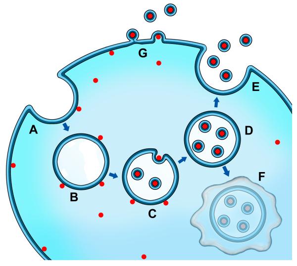
Extracellular vesicles (EVs) are released from many cell types, including normal and pathological cells, and range from 30 to 1000 nm in size. Once thought to be a mechanism for discarding unwanted cellular material, EVs are now thought to play a role in intercellular communication. Evidence is accruing that EVs are capable of carrying mRNAs, miRNAs, noncoding RNAs, and proteins, including those associated with neurodegenerative diseases and cancer, which may be exchanged between cells. For this reason, neurodegenerative diseases and cancers may share a common mechanism of disease spread via EVs. Understanding the role EVs play in disease initiation and progression will aid in the discovery of new clinically relevant biomarkers and the development of better targeted molecular and biological therapies.

Keywords: cancer; exosome; extracellular vesicles; microvesicle; neurodegeneration; stem cell.


Figures

Figure 1
Figure 1
Extracellular vesicle (EV) biogenesis and transfer. Depiction of cargo loading into EVs such as nucleic acids and intracellular proteins where endocytosis of the plasma membrane (A) results in the uptake of proteins, nucleic acids, and membrane-associated molecules (red dots), and formation of the early endosome (B). Upon transformation of the early endosome into the late endosome (C), exosomes are formed by inward budding of the late endosome/multivesicular body (MVB) with the content, including mRNA, miRNA, DNA, and protein, in a similar orientation as at the plasma membrane (D). Fusion of the MVB with the plasma membrane allows for the release of exosomes into the extracellular space (E). Alternatively, the MVB may fuse with the lysosome for degradation (F). Other EVs, such as ectosomes, can be formed directly at the plasma membrane (G). Released EVs, along with their content, may be taken up by an adjacent or distant cell.
Figure 2
Figure 2
Hypothesis of disease origin and spread by extracellular vesicles (EVs). Disease may ensue and progress by inhalation or ingestion of a virus, which could be transported to the lungs or intestinal tract (green dots). Release of EVs (small blue dots) by a virus could be then transported via the vagus nerve to brainstem nuclei, including the vagal nerve complex and then the midline raphe nuclei which connect to and interrelate synaptically-related structures of the brain (e.g. raphe and vagal nerve complex, substantia nigra, and the caudate-putamen).

References

    1. Fuxe K, et al. From the Golgi-Cajal mapping to the transmitter-based characterization of the neuronal networks leading to two modes of brain communication: wiring and volume transmission. Brain Res Rev. 2007;55:17–54. - PubMed
    1. Zhou J, et al. Predicting regional neurodegeneration from the healthy brain functional connectome. Neuron. 2012;73:1216–1227. - PMC - PubMed
    1. Pant S, et al. The multifaceted exosome: biogenesis, role in normal and aberrant cellular function, and frontiers for pharmacological and biomarker opportunities. Biochem Pharmacol. 2012;83:1484–1494. - PMC - PubMed
    1. Steindler DA, Bradley RH. N-[acetyl-3H] wheat germ agglutinin: anatomical and biochemical studies of a sensitive bidirectionally transported axonal tracer. Neuroscience. 1983;10:219–241. - PubMed
    1. Manterola L, et al. A small noncoding RNA signature found in exosomes of GBM patient serum as a diagnostic tool. Neuro Oncol. 2014;16:520–527. - PMC - PubMed

Publication types