. 2014 May 16;9(5):e97406.
doi: 10.1371/journal.pone.0097406. eCollection 2014.

Overexpression of SATB1 is associated with biologic behavior in human renal cell carcinoma

Affiliations

Overexpression of SATB1 is associated with biologic behavior in human renal cell carcinoma

Chao Cheng et al. PLoS One. .

Retraction in

Abstract

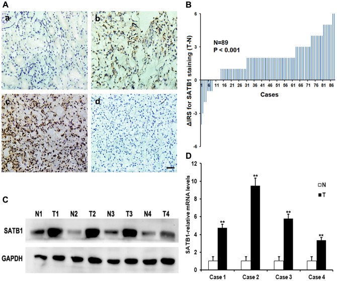
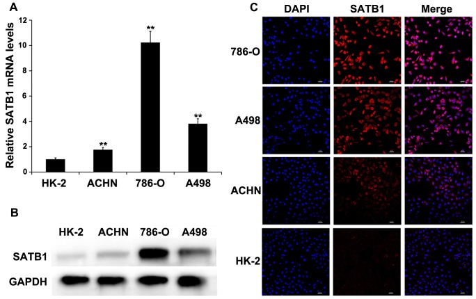
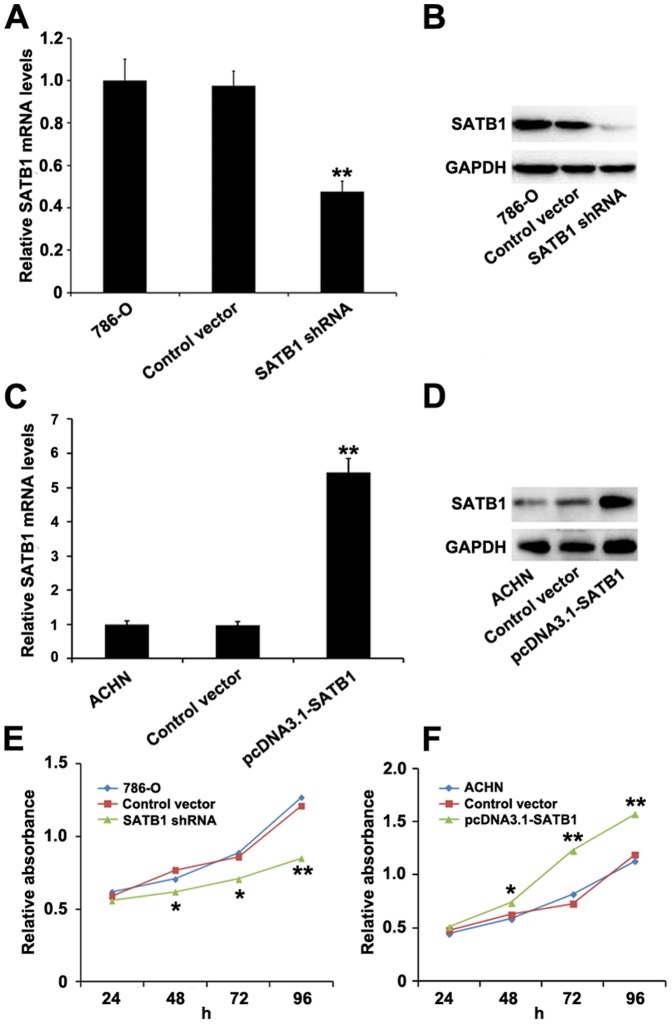
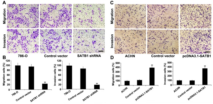
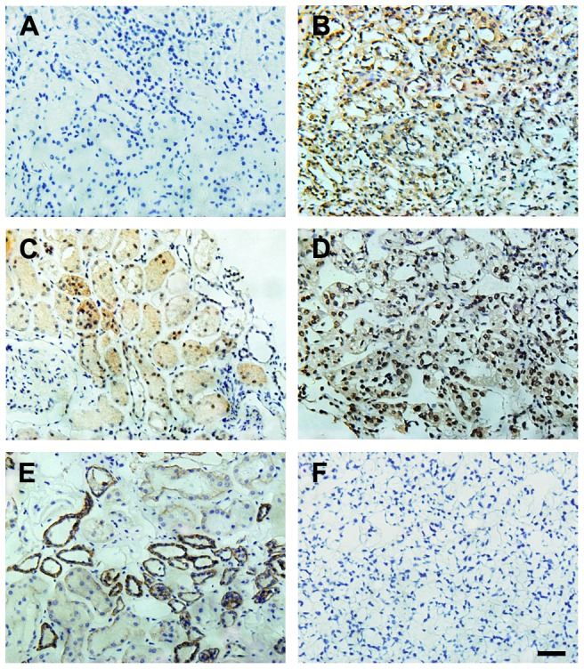
Special AT-rich sequence-binding protein-1 (SATB1) has been reported to be aberrantly expressed in various cancers and correlated with the malignant behavior of cancer cells. However, the function of SATB1 in RCC remains unclear. With the combination of immunohistochemistry, western blotting, immunofluorescence, qRT-PCR, and cell proliferation, migration and invasion assays, we found that levels of SATB1 mRNA and protein were dramatically increased in human ccRCC tissues (P<0.001 for both), and upregulation of SATB1 was significantly associated with depth of invasion (P<0.001), lymph node status (P = 0.001) and TNM stage (P = 0.009). SATB1 knockdown inhibited the proliferation, migration and invasion of 786-O cells, whereas SATB1 overexpression promoted the growth and aggressive phenotype of ACHN cells in vitro. Furthermore, SATB1 expression was positively correlated with ZEB2 expression (P = 0.013), and inversely linked to levels of SATB2 and E-cadherin (P = 0.005 and P<0.001, respectively) in ccRCC tissues. Our data provide a basis for the concept that overexpression of SATB1 may play a critical role in the acquisition of an aggressive phenotype for RCC cells through EMT, providing new insights into the significance of SATB1 in invasion and metastasis of ccRCC, which may contribute to fully elucidating the exact mechanism of development and progression of RCC.


Conflict of interest statement

Competing Interests: The authors have declared that no competing interests exist.

Figures

Figure 1
Figure 1. Expressions of SATB1 in human ccRCC tissues and matched non-cancer tissues.
A, immunohistochemical staining for SATB1 in representative sections. (a) Negative or low level staining of SATB1 in paired normal tissue; (b) Moderate staining of SATB1 in tumor tissue; (c) Strong staining in majority of ccRCC cells; (d) Negative control for SATB1 staining in ccRCC tissues. Magnification: 400×. B, The distribution of the difference in immunoreactivity score (IRS) of SATB1 staining. The IRS of SATB1 was obtained from 89 pairs of tissues, and the difference was expressed as ΔIRS = IRST-IRSN (T, tumor tissues; N, matched normal tissues). C–D, Expressions of SATB1 protein and mRNA in four representative pairs of matched normal renal tissues (N) and ccRCC tissues (T) were assessed by western blotting and qRT-PCR, respectively. Scale bar: 10 µM. Data were shown as mean ± SD (n = 3). **P<0.001.
Figure 2
Figure 2. SATB1 expression is correlated with the aggressive phenotypes of renal cancer cell lines.
SATB1 expression in immortalized normal human proximal tubule epithelial cell line HK-2 and three types of RCC cell lines (ACHN, 786-O and A498) was determined by qRT-PCR (A), western blotting (B) and immunofluorescent staining (C; Magnification: 200×), respectively. GAPDH served as a loading control. Scale bar represents 20 µM. Data were shown as mean ± SD (n = 3). **P<0.001.
Figure 3
Figure 3. Effects of suppression and overexpression of SATB1 on RCC cell line 786-O and ACHN.
The 786-O cells were transfected with pGenesil2 control vector or pGenesil2-SATB1-shRNA, while ACHN cells were treated with pcDNA3.1 control vector or pcDNA3.1-SATB1, and nontransfected 786-O and ACHN cells were used as blank controls respectively. As compared to control groups, both mRNA and protein levels of SATB1were significantly decreased in 786-O cells after SATB1 knockdown (A, B), and remarkably increased in ACHN cells with SATB1 overexpression (C, D). Evaluation of cell proliferation by CCK-8 assays showed that down-regulation of SATB1 dramatically inhibited the cell proliferation rate of 786-O cells (E), whereas overexpression of SATB1 significantly promoted the growth of ACHN cells (F). Data were shown as mean ± SD (n = 3). *P<0.05, **P<0.001.
Figure 4
Figure 4. Cell migration and invasion potential in vitro were evaluated by transwell migration and invasion assays.
A, Silencing of SATB1 inhibited the cell migration and invasion capability of 786-O cells. B, Quantitative analysis of the number of 786-O cells that successfully migrated and invaded. C, SATB1 overexpression promoted the migration and invasion of ACHN cells. D, The number of migrated and invaded ACHN cells increased with SATB1 upregulation. Scale bar: 10 µM. Data were shown as mean ± SD (n = 3). **P<0.001. Magnification: 200×.
Figure 5
Figure 5. Correlations between SATB1 and EMT markers or SATB2 in ccRCC tissues and paired control tissues.
Negative SATB1 staining (A) and high expressions of SATB2 (C) and E-cadherin (E) were shown in matched normal renal tissues, respectively. Positive SATB1 staining (B) and high ZEB2 expression (D) were observed in ccRCC tissues. (F) Negative control in ccRCC tissues. (Hematoxylin counterstain; Magnification: 400×). Scale bar: 10 µM.

References

    1. Fang Z, Tang Y, Fang J, Zhou Z, Xing Z, et al. (2013) Simvastatin inhibits renal cancer cell growth and metastasis via AKT/mTOR, ERK and JAK2/STAT3 pathway. PLoS One 8: e62823. - PMC - PubMed
    1. Xue YJ, Xiao RH, Long DZ, Zou XF, Wang XN, et al. (2012) Overexpression of FoxM1 is associated with tumor progression in patients with clear cell renal cell carcinoma. J Transl Med 10: 200. - PMC - PubMed
    1. Basso M, Cassano A, Barone C (2010) A survey of therapy for advanced renal cell carcinoma. Urol Oncol 28: 121–133. - PubMed
    1. Rini BI, Campbell SC, Escudier B (2009) Renal cell carcinoma. Lancet 373: 1119–1132. - PubMed
    1. Jiang Z, Chu PG, Woda BA, Liu Q, Balaji KC, et al. (2008) Combination of quantitative IMP3 and tumor stage: a new system to predict metastasis for patients with localized renal cell carcinomas. Clin Cancer Res 14: 5579–5584. - PubMed

Publication types

MeSH terms