. 2014 Jun 3;111(22):8055-60.
doi: 10.1073/pnas.1323788111. Epub 2014 May 16.

How cells flow in the spreading of cellular aggregates

Affiliations

How cells flow in the spreading of cellular aggregates

Grégory Beaune et al. Proc Natl Acad Sci U S A. .

Abstract

Like liquid droplets, cellular aggregates, also called "living droplets," spread onto adhesive surfaces. When deposited onto fibronectin-coated glass or polyacrylamide gels, they adhere and spread by protruding a cellular monolayer (precursor film) that expands around the droplet. The dynamics of spreading results from a balance between the pulling forces exerted by the highly motile cells at the periphery of the film, and friction forces associated with two types of cellular flows: (i) permeation, corresponding to the entry of the cells from the aggregates into the film; and (ii) slippage as the film expands. We characterize these flow fields within a spreading aggregate by using fluorescent tracking of individual cells and particle imaging velocimetry of cell populations. We find that permeation is limited to a narrow ring of width ξ (approximately a few cells) at the edge of the aggregate and regulates the dynamics of spreading. Furthermore, we find that the subsequent spreading of the monolayer depends heavily on the substrate rigidity. On rigid substrates, the migration of the cells in the monolayer is similar to the flow of a viscous liquid. By contrast, as the substrate gets softer, the film under tension becomes unstable with nucleation and growth of holes, flows are irregular, and cohesion decreases. Our results demonstrate that the mechanical properties of the environment influence the balance of forces that modulate collective cell migration, and therefore have important implications for the spreading behavior of tissues in both early development and cancer.

Keywords: tissue dynamics; tissue mechanosensitivity; wetting.


Conflict of interest statement

The authors declare no conflict of interest.

Figures

Fig. 1.
Fig. 1.
Spreading of large and small cell aggregates on glass coverslips (E = 70 GPa) coated with fibronectin and highlighted permeation process. Aggregates are observed in confocal microscopy (GFP and CHFP fluorescence). (A) Spreading of large (Upper, R0 = 185 µm) and small aggregate (Lower, R0 = 65 µm) observed at different times (t = 0–10 h). (B) Spreading of the large (Left) and small aggregate (Right) in GFP fluorescence 7 h after their deposition. Dynamics of the cells is followed using manual tracking (ImageJ). (C) The different noticeable areas of the large (Left) and small (Right) aggregates 9 h after their deposition. (D) Flows in a large (Upper) and small (Lower) cellular aggregate spreading on a substrate; the precursor film is fed by permeation (Right). The a, b, and c domains correspond to immobile cells, permeation domains, and flowing monolayers, respectively. All scale bars represent 50 μm in each panel, respectively.
Fig. 2.
Fig. 2.
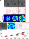
(A) Spreading of an aggregate on a coverslip (E = 70 GPa) observed in bright field at short times (Top). Using the PIV method, the direction of velocity fields (Middle) and heat maps showing the spatial distribution of velocity fields (Bottom) have been obtained. (B) Bright-field picture of the same aggregate at long times (50 h, Left). Heat map showing the spatial distribution of a velocity field (Center Left). The direction of the corresponding velocity field shows the formation of swirls (Center Right). Magnified view of the region delimited by the red box (Right). (C) Time evolution of the monolayer area of spreading aggregates normalized by the initial aggregates radius R0. Red markers (n = 8 experiments with R0 ranging from 69 to 139 µm) correspond to the case of glass coverslips and black markers (N = 8 experiments with R0 ranging from 146 to 183 µm) correspond to rigid gels (E = 16.7 kPa).
Fig. 3.
Fig. 3.
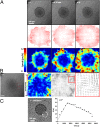
(A) Spreading of an aggregate on a PAA gel (E = 16.7 kPa) observed in bright field at short times (Top). Using the PIV method the direction of velocity fields (Middle) and heat maps show the spatial distribution of velocity fields (Bottom) have been obtained. (B) Bright-field picture of the same aggregate at long times (35 h, Left) and heat map showing spatial distribution of a velocity field (Center Left). The direction of the corresponding velocity field shows the formation of whirls (Center Right). Magnified view of the region delimited by the red box (Right). (C) Spreading of an aggregate on a PAA gel (E = 9 kPa) observed in bright field (Left). The opening and closing of a hole, circled in white on the picture, is represented by the time evolution of its radius R (Right).

References

    1. Poujade M, et al. Collective migration of an epithelial monolayer in response to a model wound. Proc Natl Acad Sci USA. 2007;104(41):15988–15993. - PMC - PubMed
    1. Tamada M, Perez TD, Nelson WJ, Sheetz MP. Two distinct modes of myosin assembly and dynamics during epithelial wound closure. J Cell Biol. 2007;176(1):27–33. - PMC - PubMed
    1. Jacinto A, Martinez-Arias A, Martin P. Mechanisms of epithelial fusion and repair. Nat Cell Biol. 2001;3(5):E117–E123. - PubMed
    1. Tambe DT, et al. Collective cell guidance by cooperative intercellular forces. Nat Mater. 2011;10(6):469–475. - PMC - PubMed
    1. Lepage SE, Bruce AEE. Zebrafish epiboly: Mechanics and mechanisms. Int J Dev Biol. 2010;54(8-9):1213–1228. - PubMed

Publication types

MeSH terms