

Longitudinal changes of angle configuration in primary angle-closure suspects: the Zhongshan Angle-Closure Prevention Trial
- PMID: 24835757
- PMCID: PMC4624262
- DOI: 10.1016/j.ophtha.2014.03.039
Longitudinal changes of angle configuration in primary angle-closure suspects: the Zhongshan Angle-Closure Prevention Trial
Abstract
Objective: To determine longitudinal changes in angle configuration in the eyes of primary angle-closure suspects (PACS) treated by laser peripheral iridotomy (LPI) and in untreated fellow eyes.
Design: Longitudinal cohort study.
Participants: Primary angle-closure suspects aged 50 to 70 years were enrolled in a randomized, controlled clinical trial.
Methods: Each participant was treated by LPI in 1 randomly selected eye, with the fellow eye serving as a control. Angle width was assessed in a masked fashion using gonioscopy and anterior segment optical coherence tomography (AS-OCT) before and at 2 weeks, 6 months, and 18 months after LPI.
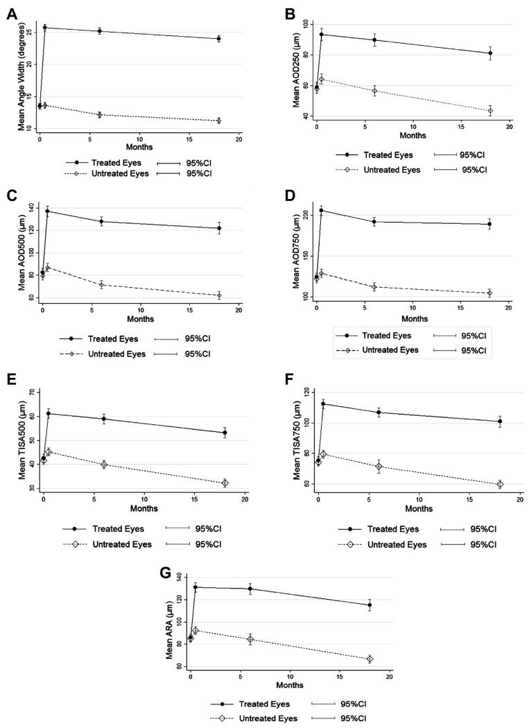
Main outcome measures: Angle width in degrees was calculated from Shaffer grades assessed under static gonioscopy. Angle configuration was also evaluated using angle opening distance (AOD250, AOD500, AOD750), trabecular-iris space area (TISA500, TISA750), and angle recess area (ARA) measured in AS-OCT images.
Results: No significant difference was found in baseline measures of angle configuration between treated and untreated eyes. At 2 weeks after LPI, the drainage angle on gonioscopy widened from a mean of 13.5° at baseline to a mean of 25.7° in treated eyes, which was also confirmed by significant increases in all AS-OCT angle width measures (P<0.001 for all variables). Between 2 weeks and 18 months after LPI, a significant decrease in angle width was observed over time in treated eyes (P<0.001 for all variables), although the change over the first 5.5 months was not statistically significant for angle width measured under gonioscopy (P = 0.18), AOD250 (P = 0.167) and ARA (P = 0.83). In untreated eyes, angle width consistently decreased across all follow-up visits after LPI, with a more rapid longitudinal decrease compared with treated eyes (P values for all variables ≤0.003). The annual rate of change in angle width was equivalent to 1.2°/year (95% confidence interval [CI], 0.8-1.6) in treated eyes and 1.6°/year (95% CI, 1.3-2.0) in untreated eyes (P<0.001).
Conclusions: Angle width of treated eyes increased markedly after LPI, remained stable for 6 months, and then decreased significantly by 18 months after LPI. Untreated eyes experienced a more consistent and rapid decrease in angle width over the same time period.
Copyright © 2014 American Academy of Ophthalmology. Published by Elsevier Inc. All rights reserved.
Figures
References
-
- Foster PJ, Oen FT, Machin D, et al. The prevalence of glaucoma in Chinese residents of Singapore: a cross-sectional population survey of the Tanjong Pagar district. Arch Ophthalmol. 2000;118:1105–11. - PubMed
-
- Foster PJ, Baasanhu J, Alsbirk PH, et al. Glaucoma in Mongolia. A population-based survey in Hovsgol province, northern Mongolia. Arch Ophthalmol. 1996;114:1235–41. - PubMed
-
- Congdon N, Wang F, Tielsch JM. Issues in the epidemiology and population-based screening of primary angle-closure glaucoma. Surv Ophthalmol. 1992;36:411–23. - PubMed
-
- He M, Foster PJ, Ge J, et al. Prevalence and clinical characteristics of glaucoma in adult Chinese: a population-based study in Liwan District, Guangzhou. Invest Ophthalmol Vis Sci. 2006;47:2782–8. - PubMed
Publication types
MeSH terms
Grants and funding
LinkOut - more resources
Full Text Sources
Other Literature Sources
Research Materials
