. 2014 Jun 10;22(6):830-41.
doi: 10.1016/j.str.2014.04.003. Epub 2014 May 15.

Multiple functional roles of the accessory I-domain of bacteriophage P22 coat protein revealed by NMR structure and CryoEM modeling

Affiliations

Multiple functional roles of the accessory I-domain of bacteriophage P22 coat protein revealed by NMR structure and CryoEM modeling

Alessandro A Rizzo et al. Structure. .

Abstract

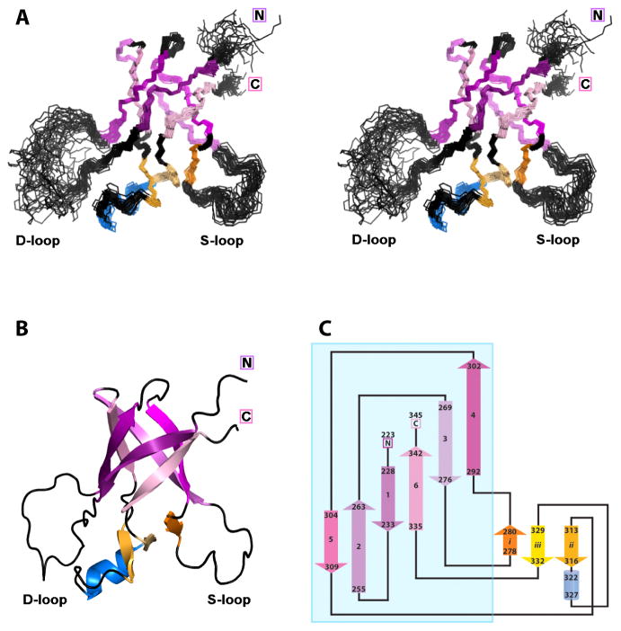
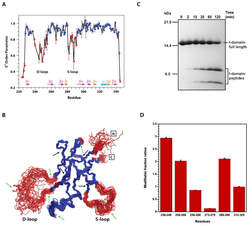
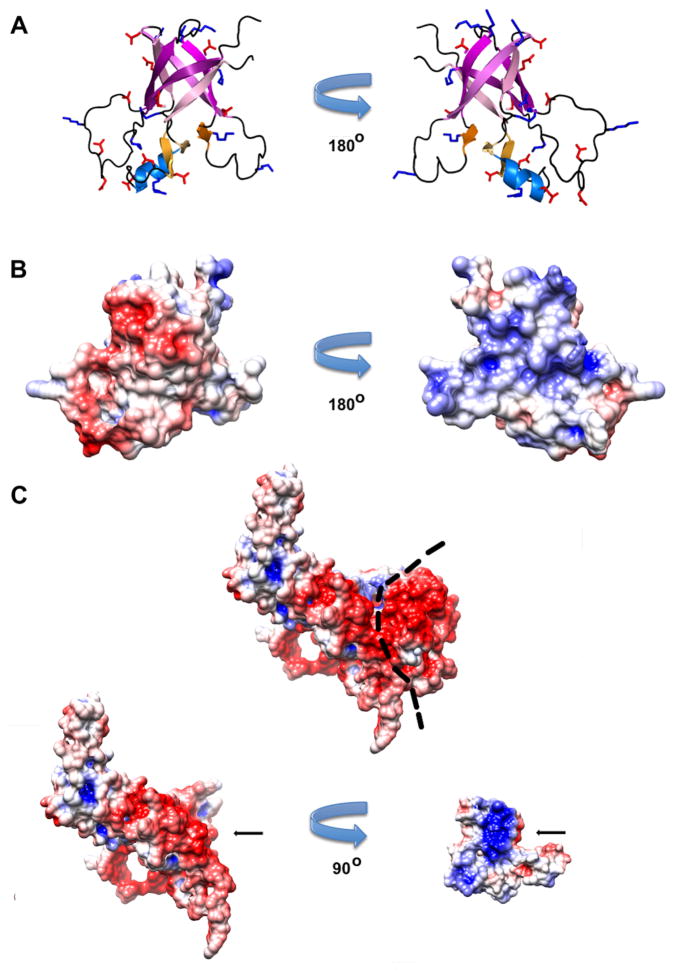
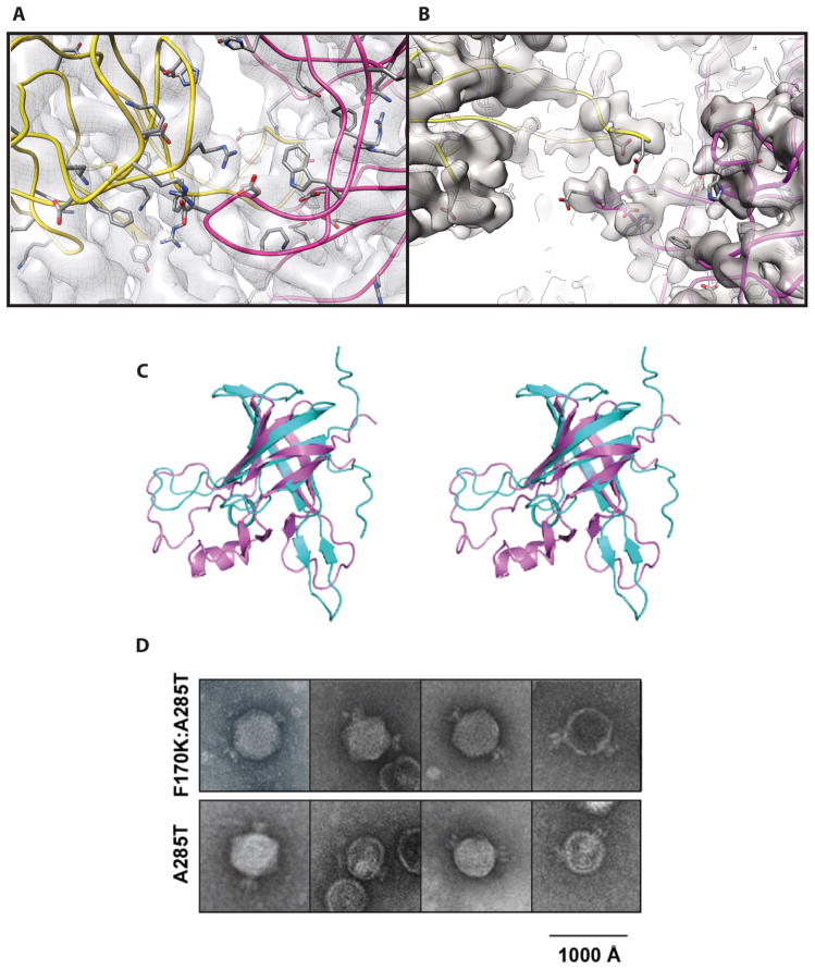
Some capsid proteins built on the ubiquitous HK97-fold have accessory domains imparting specific functions. Bacteriophage P22 coat protein has a unique insertion domain (I-domain). Two prior I-domain models from subnanometer cryoelectron microscopy (cryoEM) reconstructions differed substantially. Therefore, the I-domain's nuclear magnetic resonance structure was determined and also used to improve cryoEM models of coat protein. The I-domain has an antiparallel six-stranded β-barrel fold, not previously observed in HK97-fold accessory domains. The D-loop, which is dynamic in the isolated I-domain and intact monomeric coat protein, forms stabilizing salt bridges between adjacent capsomers in procapsids. The S-loop is important for capsid size determination, likely through intrasubunit interactions. Ten of 18 coat protein temperature-sensitive-folding substitutions are in the I-domain, indicating its importance in folding and stability. Several are found on a positively charged face of the β-barrel that anchors the I-domain to a negatively charged surface of the coat protein HK97-core.


Figures

Figure 1
Figure 1
(A) Assembly of bacteriophage P22. (B) Crystal structure of HK97 coat protein (PDB 1OHG). (C). Model of the P22 coat protein subunit from the sub-nanometer resolution cryoEM reconstruction of the mature capsid (PDB 3IYH, EMDB ID: 5150). The I-domain is shown in magenta.
Figure 2
Figure 2
NMR structure of the I-domain. (A) Stereo diagram showing the backbones of the final 30 lowest-energy NMR structures. The six strands that comprise the β-barrel are colored with a gradient going from dark purple (N-terminus) to light purple (C-terminus). Secondary structure outside the β-barrel core includes a small three-stranded β-sheet (orange) and a two-turn α-helix (light blue). (B) Ribbon diagram of the NMR structure closest to the ensemble average, with the coloring scheme described above. (C) Topology diagram of the I-domain structure, as generated with the Pro-origami server (Stivala et al., 2011). The sequence position of each secondary structure element is given in the figure. The blue box indicates the major domain.
Figure 3
Figure 3
Dynamics of the I-domain. (A) Backbone dynamics of the I-domain. S2 order parameters above 0.85 indicative of rigid structure are shown in blue; those lower than 0.85 corresponding to flexible regions are shown in red. Elements of regular secondary structure in the I-domain are indicated at the bottom of the plot. (B) NMR ensemble of the I-domain showing the agreement between the precision of the backbone in the NMR structures and S2 order parameters. The coloring scheme is the same as in (A). (C) Time course for trypsin digestion of the I-domain followed by 16% tricine SDS-PAGE. Peptides from proteolytic cleavage were analyzed by in-gel digestion and liquid chromatography coupled with tandem mass spectrometry. Arrows in (B) indicate all possible trypsin cut sites within the I-domain. The green arrows are sites where cleavage occurred, while black arrows are sites resistant to trypsin. (D) Footprinting of the I-domain in the full-length protein using FPOP and mass spectrometry. The red bars show the normalized modification levels of peptides compared to peptide 312–325.
Figure 4
Figure 4
Comparison of structural models for the I-domain. (A) Ribbon diagram of the I-domain NMR structure colored according to residue position in the sequence (N-terminus - blue, C-terminus - red). (B) Model of the I-domain structure from the 8.2 Å-resolution cryoEM reconstruction (Parent et al., 2010). The coloring scheme is identical to that in (A) but residues 314–345 (red) could not be modeled using the cryoEM data. (C) Model of the I-domain structure from the 3.8 Å-resolution cryoEM reconstruction (Chen et al., 2011). This model has only Cα atoms so secondary structure elements are not identified. The colors indicating the sequential order of segments are directly comparable to those in (A). (D) Refined P22 capsid protein, containing the I-domain NMR structure, from the 3.8 Å resolution procapsid cryoEM density map. (E) Refined P22 capsid protein, containing the I-domain NMR structure, from the 4.0 Å resolution mature virion cryoEM density map. In (D) and (E), the I-domain is rainbow colored as in 4A. The arrows point to the F-loop. (F) CryoEM density map refined model of the P22 procapsid asymmetric unit. (G) CryoEM density map refined model of the P22 virion asymmetric unit. In (F) and (G), individual P22 coat protein subunits are colored and superimposed on the respective cryoEM density maps (grey).
Figure 5
Figure 5
Distribution of charged residues in the I-domain structure. (A) Ribbon representations of the I-Domain NMR structure showing the locations of acidic (red) and basic (blue) residues on opposite faces of the β-barrel. (B) Electrostatic surface maps corresponding to the views in (A). (C) Electrostatic potential map showing the entire P22 coat protein monomer reconstructed from cryoEM data in conjunction with the I-domain NMR structure. The dotted line denotes the binding interface between the I-domain and the HK97-core of the coat protein. In the view below, the HK97-core (left) is separated from the I-domain (right). The I-domain is rotated by 90° to emphasize the patch of positive charges (blue) that interacts with the patch of negative charges (red) on the HK97-core part of the core protein. The arrows indicate how the I-domain fits into the HK97-core. Electrostatic potentials were calculated with the with the APBS server (Baker et al., 2001).
Figure 6
Figure 6
Potential functions of the I-domain. (A) A zoomed in view of the procapsid D-loops at the 2-fold from the interior. Amino acids side chains of charged residues in the D-loop are displayed to show the cluster of electrostatic interactions. Given the resolution of the model, exact locations of side chains are unclear but the region shown is highly charged. (B) Zoomed in view of the virion D-loops at the 2-fold from the interior. Note the smaller number of contacts in the mature virion (B) compared to the procapsid (A). (C) Stereo-diagram showing the least-squares superposition of the I-domain NMR structure (purple) and the RIEf fold (cyan) from S. solfataricus translation initiation factor 2γ (PDB code 3P3M, chain A, residues 205–325). (D) Negative stain electron micrographs of capsids assembled from coat protein with the A285T or F170K:A285T substitutions. The mutant phage particles exhibit multiple tails, indicating the portal complex is incorporated at more than one vertex.

References

    1. Abrescia NG, Bamford DH, Grimes JM, Stuart DI. Structure unifies the viral universe. Annual Review of Biochemistry. 2012;81:795–822. - PubMed
    1. Adams PD, Afonine PV, Bunkoczi G, Chen VB, Echols N, Headd JJ, Hung LW, Jain S, Kapral GJ, Grosse Kunstleve RW, et al. The Phenix software for automated determination of macromolecular structures. Methods. 2011;55:94–106. - PMC - PubMed
    1. Akita F, Chong KT, Tanaka H, Yamashita E, Miyazaki N, Nakaishi Y, Suzuki M, Namba K, Ono Y, Tsukihara T, et al. The crystal structure of a virus-like particle from the hyperthermophilic archaeon Pyrococcus furiosus provides insight into the evolution of viruses. Journal of Molecular Biology. 2007;368:1469–1483. - PubMed
    1. Alexandrescu AT, Jahnke W, Wiltscheck R, Blommers MJ. Accretion of structure in staphylococcal nuclease: an 15N NMR relaxation study. Journal of molecular biology. 1996;260:570–587. - PubMed
    1. Baker ML, Hryc CF, Zhang Q, Wu W, Jakana J, Haase-Pettingell C, Afonine PV, Adams PD, King JA, Jiang W, et al. Validated near-atomic resolution structure of bacteriophage epsilon15 derived from cryo-EM and modeling. Proc Natl Acad Sci U S A. 2013;110:12301–12306. - PMC - PubMed

Publication types

MeSH terms

LinkOut - more resources