

Active invasion of oral and aortic tissues by Porphyromonas gingivalis in mice causally links periodontitis and atherosclerosis
- PMID: 24836175
- PMCID: PMC4024021
- DOI: 10.1371/journal.pone.0097811
Active invasion of oral and aortic tissues by Porphyromonas gingivalis in mice causally links periodontitis and atherosclerosis
Abstract
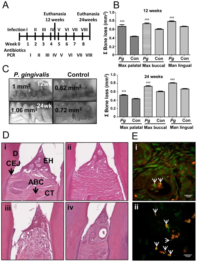
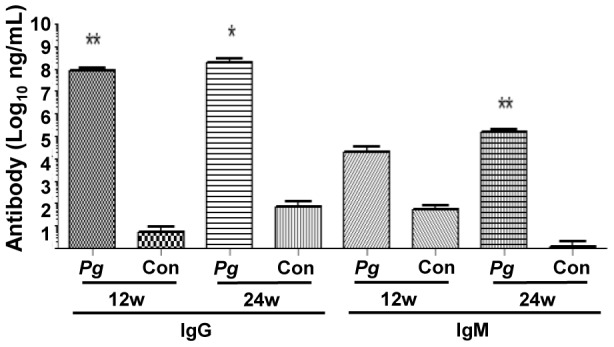
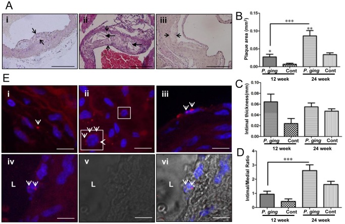
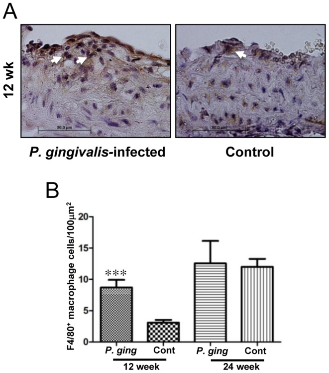
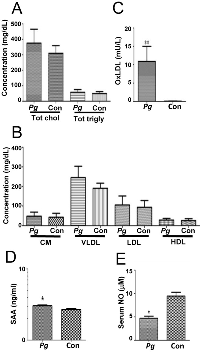
Atherosclerotic vascular disease is a leading cause of myocardial infarction and cerebrovascular accident, and independent associations with periodontal disease (PD) are reported. PD is caused by polymicrobial infections and aggressive immune responses. Genomic DNA of Porphyromonas gingivalis, the best-studied bacterial pathogen associated with severe PD, is detected within atherosclerotic plaque. We examined causal relationships between chronic P. gingivalis oral infection, PD, and atherosclerosis in hyperlipidemic ApoEnull mice. ApoEnull mice (n = 24) were orally infected with P. gingivalis for 12 and 24 weeks. PD was assessed by standard clinical measurements while the aorta was examined for atherosclerotic lesions and inflammatory markers by array. Systemic inflammatory markers serum amyloid A, nitric oxide, and oxidized low-density lipoprotein were analyzed. P. gingivalis infection elicited specific antibodies and alveolar bone loss. Fluorescent in situ hybridization detected viable P. gingivalis within oral epithelium and aorta, and genomic DNA was detected within systemic organs. Aortic plaque area was significantly increased in P. gingivalis-infected mice at 24 weeks (P<0.01). Aortic RNA and protein arrays indicated a strong Th2 response. Chronic oral infection with P. gingivalis results in a specific immune response, significant increases in oral bone resorption, aortic inflammation, viable bacteria in oral epithelium and aorta, and plaque development.
Conflict of interest statement
Figures
Similar articles
-
Periodontal pathogens invade gingiva and aortic adventitia and elicit inflammasome activation in αvβ6 integrin-deficient mice.Infect Immun. 2015 Dec;83(12):4582-93. doi: 10.1128/IAI.01077-15. Epub 2015 Sep 14. Infect Immun. 2015. PMID: 26371120 Free PMC article.
-
Polymicrobial infection with major periodontal pathogens induced periodontal disease and aortic atherosclerosis in hyperlipidemic ApoE(null) mice.PLoS One. 2013;8(2):e57178. doi: 10.1371/journal.pone.0057178. Epub 2013 Feb 25. PLoS One. 2013. PMID: 23451182 Free PMC article.
-
Fusobacterium nucleatum Alters Atherosclerosis Risk Factors and Enhances Inflammatory Markers with an Atheroprotective Immune Response in ApoE(null) Mice.PLoS One. 2015 Jun 16;10(6):e0129795. doi: 10.1371/journal.pone.0129795. eCollection 2015. PLoS One. 2015. PMID: 26079509 Free PMC article.
-
Porphyromonas gingivalis mediated periodontal disease and atherosclerosis: disparate diseases with commonalities in pathogenesis through TLRs.Curr Pharm Des. 2007;13(36):3665-75. doi: 10.2174/138161207783018554. Curr Pharm Des. 2007. PMID: 18220804 Review.
-
Review: Pathogen-induced inflammation at sites distant from oral infection: bacterial persistence and induction of cell-specific innate immune inflammatory pathways.Mol Oral Microbiol. 2010 Oct;25(5):305-16. doi: 10.1111/j.2041-1014.2010.00582.x. Mol Oral Microbiol. 2010. PMID: 20883220 Free PMC article. Review.
Cited by
-
Connection between Periodontitis-Induced Low-Grade Endotoxemia and Systemic Diseases: Neutrophils as Protagonists and Targets.Int J Mol Sci. 2021 Apr 28;22(9):4647. doi: 10.3390/ijms22094647. Int J Mol Sci. 2021. PMID: 33925019 Free PMC article. Review.
-
Porphyromonas gingivalis outside the oral cavity.Odontology. 2022 Jan;110(1):1-19. doi: 10.1007/s10266-021-00647-8. Epub 2021 Aug 19. Odontology. 2022. PMID: 34410562 Review.
-
Polymicrobial Oral Infection with Four Periodontal Bacteria Orchestrates a Distinct Inflammatory Response and Atherosclerosis in ApoE null Mice.PLoS One. 2015 Nov 30;10(11):e0143291. doi: 10.1371/journal.pone.0143291. eCollection 2015. PLoS One. 2015. PMID: 26619277 Free PMC article.
-
Pseudomonas aeruginosa Microcolonies in Coronary Thrombi from Patients with ST-Segment Elevation Myocardial Infarction.PLoS One. 2016 Dec 28;11(12):e0168771. doi: 10.1371/journal.pone.0168771. eCollection 2016. PLoS One. 2016. PMID: 28030624 Free PMC article.
-
Infection of Porphyromonas gingivalis Increases Phosphate-Induced Calcification of Vascular Smooth Muscle Cells.Cells. 2020 Dec 15;9(12):2694. doi: 10.3390/cells9122694. Cells. 2020. PMID: 33334022 Free PMC article.
References
-
- Lockhart PB, Bolger AF, Papapanou PN, Osinbowale O, Trevisan M, et al. (2012) Periodontal Disease and Atherosclerotic Vascular Disease: Does the Evidence Support an Independent Association? A Scientific Statement From the American Heart Association. Circulation 125: 2520–2544 10.1161/CIR.0b013e31825719f3 - DOI - PubMed
-
- Paquette DW, Brodala N, Nichols TC (2007) Cardiovascular disease, inflammation, and periodontal infection. Periodontol 2000 44: 113–126. - PubMed
Publication types
MeSH terms
Grants and funding
LinkOut - more resources
Full Text Sources
Other Literature Sources
Medical
Molecular Biology Databases
