. 2015 Sep;25(9):3025-35.
doi: 10.1093/cercor/bhu098. Epub 2014 May 16.

Rapid Bidirectional Reorganization of Cortical Microcircuits

Affiliations

Rapid Bidirectional Reorganization of Cortical Microcircuits

Giorgia Albieri et al. Cereb Cortex. 2015 Sep.

Abstract

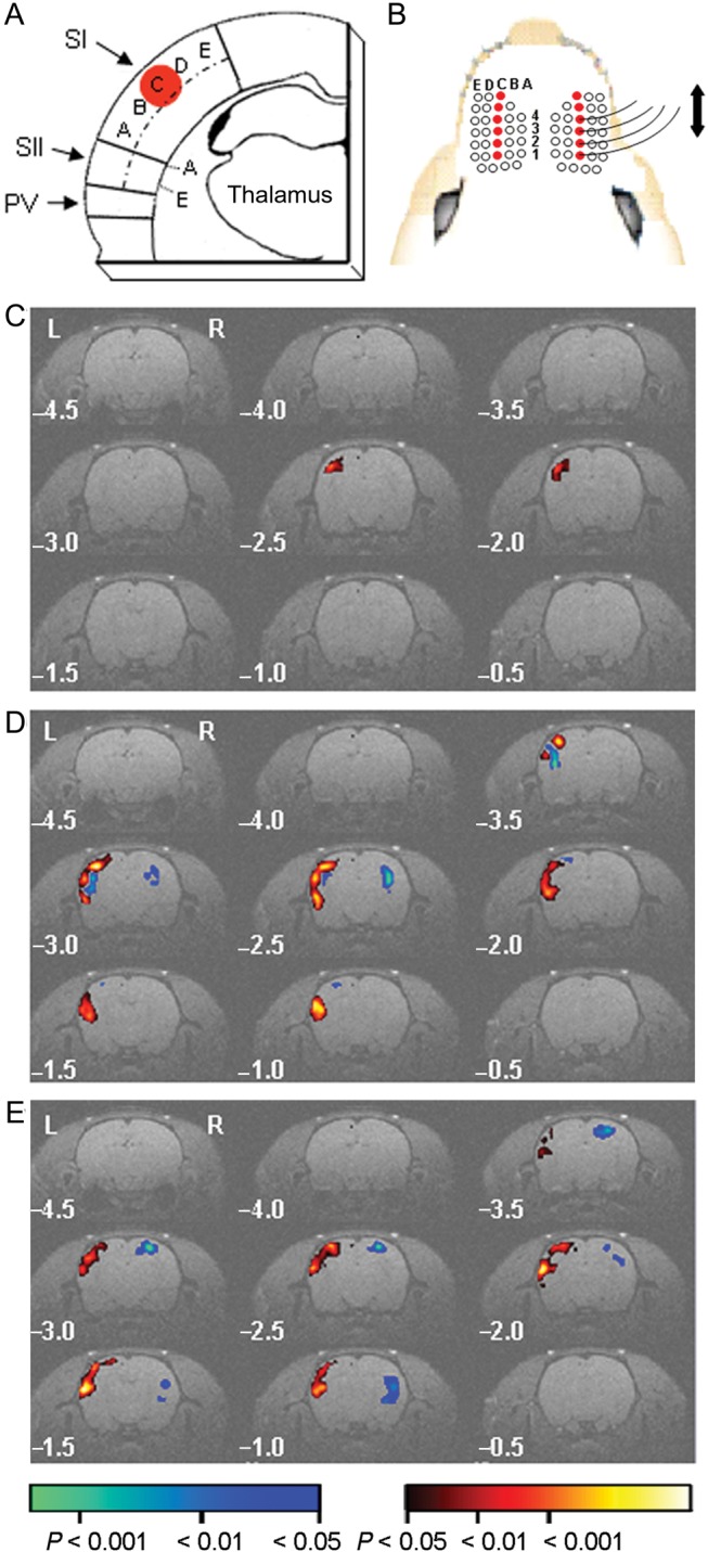
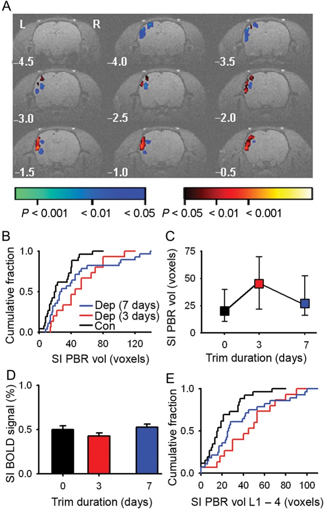
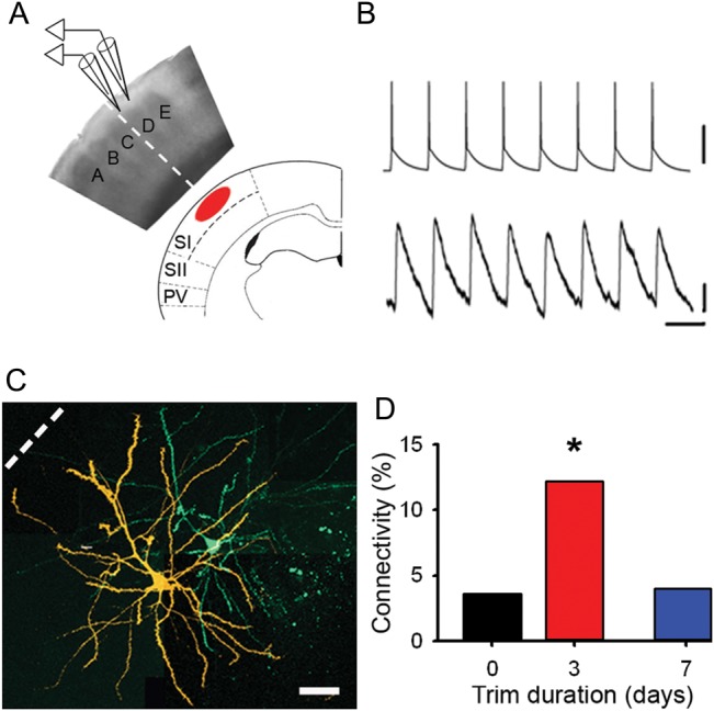
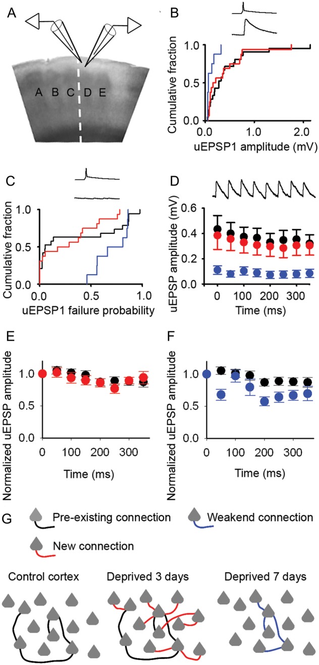
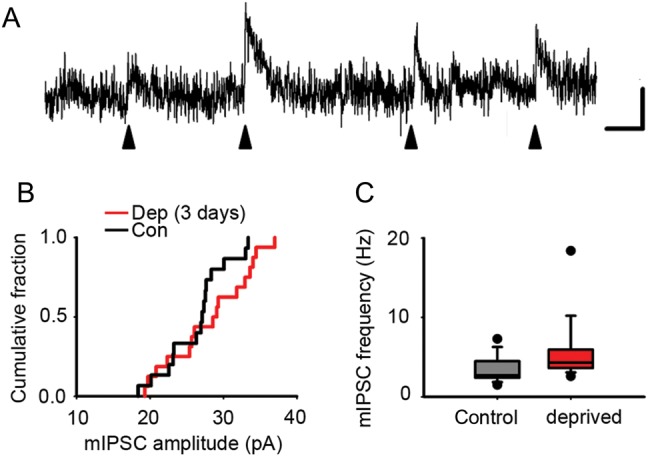
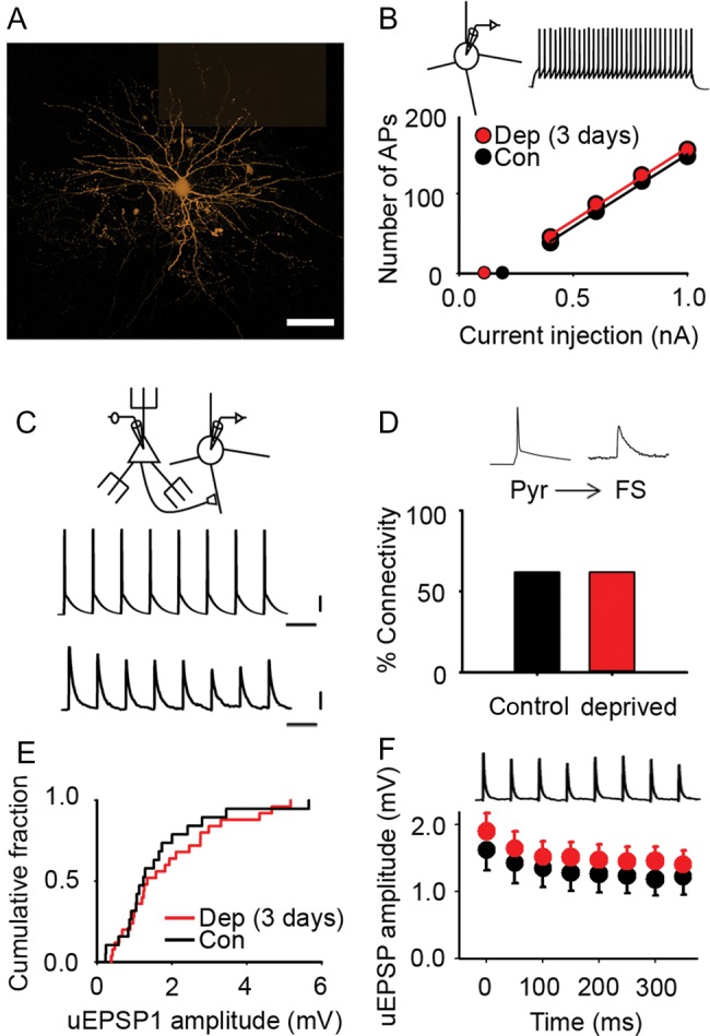
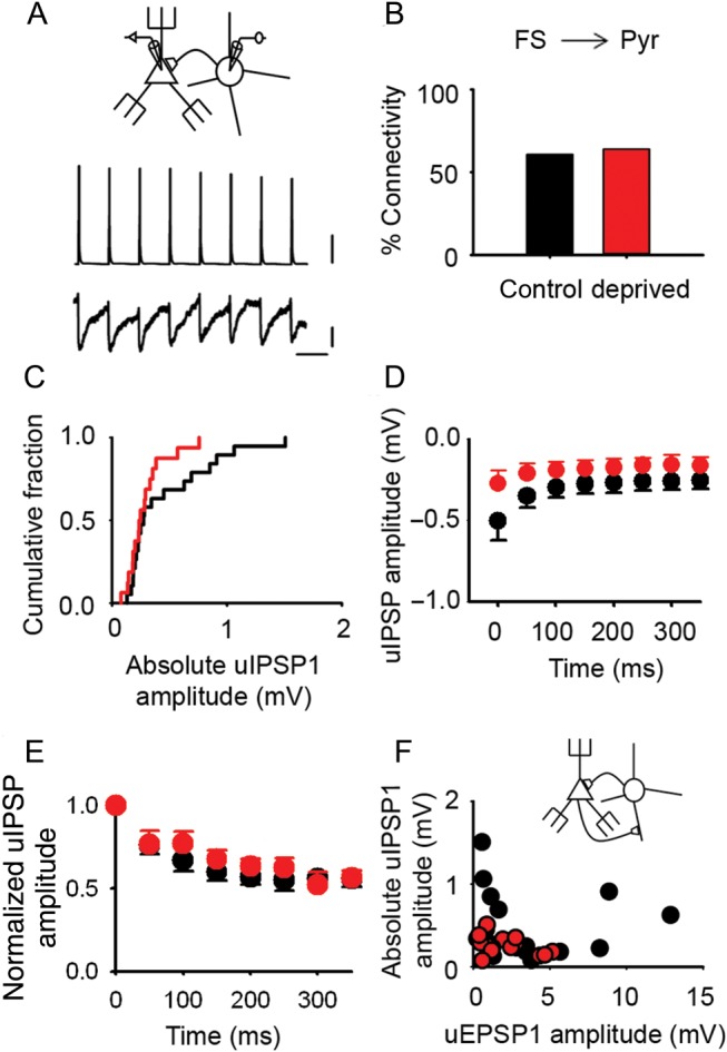
Mature neocortex adapts to altered sensory input by changing neural activity in cortical circuits. The underlying cellular mechanisms remain unclear. We used blood oxygen level-dependent (BOLD) functional magnetic resonance imaging (fMRI) to show reorganization in somatosensory cortex elicited by altered whisker sensory input. We found that there was rapid expansion followed by retraction of whisker cortical maps. The cellular basis for the reorganization in primary somatosensory cortex was investigated with paired electrophysiological recordings in the periphery of the expanded whisker representation. During map expansion, the chance of finding a monosynaptic connection between pairs of pyramidal neurons increased 3-fold. Despite the rapid increase in local excitatory connectivity, the average strength and synaptic dynamics did not change, which suggests that new excitatory connections rapidly acquire the properties of established excitatory connections. During map retraction, entire excitatory connections between pyramidal neurons were lost. In contrast, connectivity between pyramidal neurons and fast spiking interneurons was unchanged. Hence, the changes in local excitatory connectivity did not occur in all circuits involving pyramidal neurons. Our data show that pyramidal neurons are recruited to and eliminated from local excitatory networks over days. These findings suggest that the local excitatory connectome is dynamic in mature neocortex.

Keywords: connectome; cortical microcircuit; experience-dependent plasticity; fMRI; inhibition; rewiring.


Figures

Figure 1.
Figure 1.
Spared whisker representations enlarge after whisker trimming. (A) Schematic illustrating the relative position of SI, SII, and the parietal ventral area (PV) in a coronal slice through whisker barrel cortex. Dashed line bisects SI and SII. Whisker barrel columns are marked AE. Red circle, SI PBR evoked by whisker deflection. (B) Schematic of trimming protocol (open circle denotes trimmed whisker) and deflection of right C1–4 whiskers. Left C1–4 whiskers not shown. (C and E) Group statistical parametric maps of BOLD responses evoked by 5 Hz whisker deflection in sham-trimmed rats (C, n = 26 rats), and after whisker trimming for 3 days (D, n = 15 rats) and 7 days (E, n = 28 rats). Pseudocolored voxels have a positive (red) or negative (blue) BOLD signal that is significantly different from baseline. Pseudocolor scale bar applies to (C and E). Numbers indicate rostro-caudal distance from bregma.
Figure 2.
Figure 2.
SI whisker representation expands without an increase in BOLD signal amplitude. (A) Single-animal statistical parametric map of the BOLD signal evoked by 5 Hz deflection of the right C1–4 whiskers after 3 days of whisker trimming. (B) Cumulative fraction plot of SI PBR volume from single-animal maps for controls (black, n = 26 rats), 3-day trim (red, n = 15 rats), and 7-day trim (blue, n = 28 rats). (C) Median SI PBR volume and interquartile range (error bars) after 3 and 7 days of whisker trimming (median SI PBR volume: controls, 20 [11–40] voxels, n = 26 rats; 3-day trim, 45 [24–69] voxels; n = 15 rats; 7-day trim, median volume, 26 [17–51] voxels, n = 28 rats). (D) Peak amplitude of BOLD signal (error bars, SEM) after whisker trimming (control, +0.50 ± 0.04%, n = 26 rats; 3-day trim, +0.43 ± 0.04%, n = 15 rats; 7-day trim, +0.53 ± 0.04%, n = 28 rats). (E) Cumulative fraction plot of the SI PBR volume in L1–4. Color code for (CE) as (B).
Figure 3.
Figure 3.
Local excitatory connectivity changes in concert with BOLD whisker representations. (A) Schematic showing orientation of a brain slice with respect to BOLD fMRI images. Electrophysiological recordings were made in L2/3. Dashed line indicates the boundary between spared C-row whiskers and deprived cortex. (B) Synaptically connected pyramidal neurons. Upper trace, train of action potentials in presynaptic neuron. Lower trace, evoked response in postsynaptic neuron. Scale bars: 50 mV (upper), 0.1 mV (lower); 50 ms. (C) Confocal reconstruction of the presynaptic (green) and postsynaptic (orange) pyramidal neurons. Scale bar, 50 μm. (D) Connectivity between deprived L2/3 pyramidal neurons in controls (black, 3.6%), after whisker trimming for 2–4 days (red, 12.0%) and after whisker trimming for 6–8 days (blue, 4.0%).
Figure 4.
Figure 4.
New excitatory connections in deprived cortex have similar properties to control connections. (A) Schematic of recordings from pairs of synaptically connected L2/3 pyramidal neurons. (B) Mean uEPSP amplitudes in control cortex (black) and deprived cortex after 2–4 days (red) or 6–8 days (blue) of whisker trimming (2- to 4-day trim, 0.38 ± 0.11 mV, n = 16; 6- to 8-day trim, 0.11 ± 0.03 mV, n = 8; control, 0.43 ± 0.11 mV, n = 21). Inset: presynaptic action potential, postsynaptic EPSP. (C) Failure rates of neurotransmission between pyramidal neurons after 2–4 days (red) and 6–8 days (blue) of whisker trimming (median [IQR]: 2- to 4-day trim, 0.23 [0.00–0.49], n = 16; 6- to 8-day trim, 0.74 [0.56–0.85], n = 8; control, 0.05 [0.02–0.79], n = 19). Inset: presynaptic action potential, no postsynaptic response. (D) uEPSP amplitudes during a 20-Hz stimulus train in control cortex (filled circles) or deprived cortex after 2–4 days (red) and 6–8 days (blue) of whisker trimming. Error bars, SEM. Inset: 20 Hz train of postsynaptic EPSPs. (E) Normalized uEPSP amplitudes of Pyr → Pyr connections during a 20-Hz train in deprived cortex after 2–4 days trimming (red, n = 16) and in control cortex (black, n = 21). Error bars, SEM. (F) Normalized uEPSP amplitudes of Pyr → Pyr connections during a 20-Hz train in deprived cortex after 6–8 days trimming (blue, n = 8) and in control cortex (black, n = 21). Error bars, SEM. (G) Schematic shows sparse local connectivity in control cortex prior to whisker trimming (left panel). Whisker trimming for 3 days induces a rapid 3-fold increase in local excitatory connectivity (middle panel; new connections, red). Connectivity returns to control levels following 7 days of whisker trimming (right panel; 7-day trim connections, blue).
Figure 5.
Figure 5.
Inhibitory drive onto L2/3 pyramidal cells is not decreased by 3-day whisker deprivation. (A) Example trace of mIPSCs (filled arrow heads) in an L2/3 pyramidal neuron. Scale bar: 10 pA, 20 ms. (B) Cumulative fraction of mean mIPSC amplitude recorded from L2/3 pyramidal neurons in deprived (red) and control (black) cortex (grand mean rather than mIPSC amplitudes: deprived, 28.1 ± 1.4 pA, n = 16 neurons; control, 26.3 ± 1.1 pA, n = 15 neurons). (C) mIPSC frequency recorded from L2/3 pyramidal neurons in deprived (red) and control (black) cortex (mean of mean mIPSC frequencies: deprived, 4.3 [3.6–5.9] Hz, n = 16 neurons; control, 2.7 [2.4–4.5] Hz, n = 15 neurons).
Figure 6.
Figure 6.
Excitatory transmission onto L2/3 FS interneurons in deprived cortex is not affected by brief sensory deprivation. (A) Montage of maximum intensity projections from confocal z-stacks of an L2/3 FS interneuron filled with AF568 (orange). Scale bar, 40 μm. (B) Mean number of action potentials recorded in L2/3 FS interneurons evoked by 500 ms depolarizing current pulses in control (black) and deprived (red) cortex after 2–3 days of sensory deprivation. Inset: example trace of action potentials in an L2/3 FS interneuron evoked by a +0.4-nA current pulse (500 ms). Slope of the input–output curve: deprived, 182 ± 9 action potential nA−1, n = 26 FS interneurons; control, 186 ± 10 AP nA−1, n = 32 FS interneurons. Rheobase: deprived, 0.11 ± 0.04 nA, n = 26 FS interneurons; control, 0.19 ± 0.03 nA, n = 32 FS interneurons. (C) Pyr → FS connection: 20 Hz train of action potentials in the presynaptic pyramidal neuron evokes short latency uEPSPs (average 50 trials) in the postsynaptic FS interneuron. Scale bars: 20 mV (top), 0.5 mV (bottom), 50 ms. (D) Percentage of pyramidal cell to FS interneuron pairs (Pyr → FS) that were synaptically connected in control (black, 62%) and deprived (red, 62%) cortex. (E) Empirical distribution plots of mean uEPSP1 amplitudes in deprived (red) and control (black) cortex. Median [IQR] of mean uEPSP1 amplitudes for Pyr → FS connections: deprived, 1.34 [0.93–2.76] mV, n = 25; control, 1.24 [0.88–2.00] mV; n = 19. (F) Mean uEPSP amplitude during 20 Hz trains in deprived (red, n = 25 Pyr → FS connections) and control (black, n = 19 Pyr → FS connections) cortex. Error bars, SEM.
Figure 7.
Figure 7.
Inhibitory transmission onto pyramidal neurons in L2/3 of deprived cortex is unaltered by short periods of whisker deprivation. (A) Schematic showing an FS interneuron synaptically connected to a pyramidal cell (top); train of 8 action potentials in the presynaptic FS interneuron generates 8 short latency uIPSPs in the postsynaptic pyramidal neuron (average of 50 trials). Scale bars (top to bottom): 20 mV, 0.1 mV, 100 ms. (B) Percentage of tested FS interneuron to pyramidal cell pairs (FS → Pyr) that were synaptically connected in control (black, 62%) and deprived (red, 64%) cortex. (C) Empirical distribution plots of the amplitudes of mean uIPSP1 (absolute value) in deprived (red) and control (black) cortex. Median [IQR] of mean uIPSP1 amplitudes: deprived: −0.24 [−0.35 to −0.18] mV, n = 16 FS → Pyr connections; control, −0.27 [−0.69 to −0.20] mV, n = 19 FS → Pyr connections. (D) Mean uIPSP amplitude during 20 Hz trains in deprived (red, n = 8 FS → Pyr connections) and control (black, n = 13 FS → Pyr connections) cortex. Error bars, SEM. (E) uIPSP amplitudes during a 20-Hz train normalized to uIPSP1 for each L2/3 FS → Pyr connection in 3-day deprived cortex (red, n = 8) and in control cortex (black, n = 13). Error bars are within the majority of circles. (F) Relationship between mean uEPSP1 amplitude and mean uIPSP1 amplitude (absolute values) for pairs of reciprocally connected FS interneurons and pyramidal cells in control (black) and deprived (red) cortex (correlation: deprived, r = −0.40, n = 14 reciprocally connected FS → Pyr pairs; control, r = −0.06, n = 17 reciprocally connected FS → Pyr pairs).

References

    1. Alonso BD, Lowe AS, Dear JP, Lee KC, Williams SC, Finnerty GT. 2008. Sensory inputs from whisking movements modify cortical whisker maps visualized with functional magnetic resonance imaging. Cereb Cortex. 18:1314–1325. - PMC - PubMed
    1. Antonini A, Gillespie DC, Crair MC, Stryker MP. 1998. Morphology of single geniculocortical afferents and functional recovery of the visual cortex after reverse monocular deprivation in the kitten. J Neurosci. 18:9896–9909. - PMC - PubMed
    1. Barnes SJ, Finnerty GT. 2010. Sensory experience and cortical rewiring. Neuroscientist. 16:186–198. - PubMed
    1. Benison AM, Rector DM, Barth DS. 2007. Hemispheric mapping of secondary somatosensory cortex in the rat. J Neurophysiol. 97:200–207. - PubMed
    1. Campanac E, Gasselin C, Baude A, Rama S, Ankri N, Debanne D. 2013. Enhanced intrinsic excitability in basket cells maintains excitatory-inhibitory balance in hippocampal circuits. Neuron. 77:712–722. - PubMed

Publication types

MeSH terms