. 2014 Aug;196(15):2798-806.
doi: 10.1128/JB.00069-14. Epub 2014 May 16.

Dynamics of expression and maturation of the type III secretion system of enteropathogenic Escherichia coli

Affiliations

Dynamics of expression and maturation of the type III secretion system of enteropathogenic Escherichia coli

Gal Yerushalmi et al. J Bacteriol. 2014 Aug.

Abstract

Enteropathogenic Escherichia coli (EPEC) is a major cause of food poisoning, leading to significant morbidity and mortality. EPEC virulence is dependent on a type III secretion system (T3SS), a molecular syringe employed by EPEC to inject effector proteins into host cells. The injected effector proteins subvert host cellular functions to the benefit of the infecting bacteria. The T3SS and related genes reside in several operons clustered in the locus of enterocyte effacement (LEE). We carried out simultaneous analysis of the expression dynamics of all the LEE promoters and the rate of maturation of the T3SS. The results showed that expression of the LEE1 operon is activated immediately upon shifting the culture to inducing conditions, while expression of other LEE promoters is activated only ∼70 min postinduction. Parallel analysis showed that the T3SS becomes functional around 100 min postinduction. The T3SS core proteins EscS, EscT, EscU, and EscR are predicted to be involved in the first step of T3SS assembly and are therefore included among the LEE1 genes. However, interfering with the temporal regulation of EscS, EscT, EscU, and EscR expression has only a marginal effect on the rate of the T3SS assembly. This study provides a comprehensive description of the transcription dynamics of all the LEE genes and correlates it to that of T3SS biogenesis.


Figures

FIG 1
FIG 1
Systematic identification of the LEE promoters. (A) LEE intergenic regions (IRs) larger than 40 bp (gray arrows) were cloned as transcriptional fusions upstream of the GFP reporter gene. (B) EPEC (blue) or EPEC ler::kan (green) harboring the GFP gene fusions were analyzed for green fluorescence, representing promoter activity from the sequence fused to the GFP gene. The sequences IRLEE6, IRLEE1, IRrorf3, IRLEE7, IRLEE2, IRLEE3, IRcesF, IRmap, IRLEE5, IRcesT, IRescD, and IRLEE4 were found to contain promoters, while the sequences IRrorf1, IRgrlA, IRespA, and IRespF exhibited no promoter activity, similarly to the vector. The transcription from all the promoters except PLEE1 was found to be activated by Ler. The experiment was performed at least twice for each strain, with similar results. (C) Map of the LEE promoters based on the results in panel B.
FIG 2
FIG 2
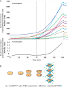
Dynamics of transcription of the LEE genes and of T3SS activity. (A) Upper panel, HeLa cells were infected with EPEC harboring the GFP gene fusion, each representing one of the LEE promoters. The level of green fluorescence was measured every 5 min, with t = 0 the time of shifting the culture to inducing conditions and initiation of infection. Lower panel, T3SS activity was measured simultaneously using HeLa cells loaded with CCF2/AM and infected with bacteria expressing a Tir-BlaM fusion. CCF2/AM hydrolysis by translocated Tir-BlaM was measured over time. Dashed vertical lines represent the time of initiation of transcription of all the LEE promoters (except PLEE1) and the time of initiation of translocation. The experiment was performed twice for each strain with similar results. (B) Schematic representation of EPEC growth, expression of LEE proteins, assembly of the T3SS, and secretion of effector proteins.
FIG 3
FIG 3
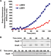
Dynamics of LEE1, LEE2, and LEE4 expression and T3SS activity. To corroborate the results shown in Fig. 2, EPEC cultures were shifted from repressive to inducing conditions and different parameters were determined. (A) Transcription from the LEE1 and LEE2 promoters was monitored using chromosomal transcriptional fusions of the GFP reporter gene to PLEE1 (strain GY2455) and PLEE2 (strain GY2529). (B) Samples of the activated bacteria were taken at the stated times postactivation and subjected to immunoblotting with anti-EspB, representing LEE4 expression, and anti-DnaK (loading control). (C) Secretion of EspB by the activated EPEC, used as readout for T3SS activity, was monitored by sampling the supernatant at the stated times postinduction and immunoblot analysis with anti-EspB. The experiment was performed twice for each strain with similar results.
FIG 4
FIG 4
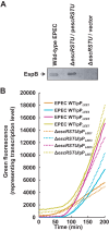
Deletion of escRSTU does not affect the kinetics of transcription of the LEE genes. (A) An EPEC escRSTU::kan strain was generated and was found to be unable to secrete EspB. The deletion was complemented by a plasmid encoding escRSTU (pGY4701) but not by the empty vector pSA10. (B) HeLa cells were infected with EPEC or EPEC escRSTU::kan harboring the GFP gene fusions with PLEE1, PLEE3, PLEE5, and PLEE7. The level of green fluorescence was measured every 5 min, with t = 0 the time of shifting the culture to inducing conditions and initiation of infection. The kinetics of the LEE gene transcription in the escRSTU::kan strain is similar to that in wild-type EPEC (WT).
FIG 5
FIG 5
Influence of delayed expression of escRSTU on T3SS activity. (A) Schematic presentation of the LEE1 and LEE6 operons of wild-type EPEC or the EPEC GY4714 strain (EPEC PLEE6-escRSTU). In GY4714, the escRSTU genes were deleted from the LEE1, replaced by a Kanr cassette, and placed in LEE6 under the regulation of the LEE6 promoter. (B to D) Cultures of wild-type EPEC and the GY4714 strain were grown under repressive conditions and shifted to inducing conditions. Three readouts for T3SS activity were compared between the two cultures: secretion of EspB (B), formation of actin pedestals (C), and cleavage of JNK (D). (B) Secretion of EspB was measured by sampling of the supernatant at the stated times postinduction and analysis by Western blotting using anti-EspB antibody. The experiment was performed twice with similar results. (C) The level of pedestal formation by infected HeLa cells was determined at different time points postinfection. Bars represent standard deviations. Results from one experiment out of two are shown. (D) Levels of JNK cleavage were determined at different time points postinfection. The arrow indicates the JNK cleavage product. The results of one experiment out of three with similar results are shown.

Similar articles

Cited by

References

    1. Nataro JP, Kaper JB. 1998. Diarrheagenic Escherichia coli. Clin. Microbiol. Rev. 11:142–201 - PMC - PubMed
    1. Lai Y, Rosenshine I, Leong JM, Frankel G. 2013. Intimate host attachment: enteropathogenic and enterohaemorrhagic Escherichia coli. Cell. Microbiol. 15:1796–1808 - PMC - PubMed
    1. Wong AR, Pearson JS, Bright MD, Munera D, Robinson KS, Lee SF, Frankel G, Hartland EL. 2011. Enteropathogenic and enterohaemorrhagic Escherichia coli: even more subversive elements. Mol. Microbiol. 80:1420–1438. 10.1111/j.1365-2958.2011.07661.x - DOI - PubMed
    1. Wagner S, Konigsmaier L, Lara-Tejero M, Lefebre M, Marlovits TC, Galan JE. 2010. Organization and coordinated assembly of the type III secretion export apparatus. Proc. Natl. Acad. Sci. U. S. A. 107:17745–17750. 10.1073/pnas.1008053107 - DOI - PMC - PubMed
    1. McDaniel TK, Kaper JB. 1997. A cloned pathogenicity island from enteropathogenic Escherichia coli confers the attaching and effacing phenotype on E. coli K-12. Mol. Microbiol. 23:399–407. 10.1046/j.1365-2958.1997.2311591.x - DOI - PubMed

Publication types

MeSH terms

LinkOut - more resources