. 2014 Jun;124(6):2599-610.
doi: 10.1172/JCI62059. Epub 2014 May 16.

β-Catenin-regulated myeloid cell adhesion and migration determine wound healing

β-Catenin-regulated myeloid cell adhesion and migration determine wound healing

Saeid Amini-Nik et al. J Clin Invest. 2014 Jun.

Abstract

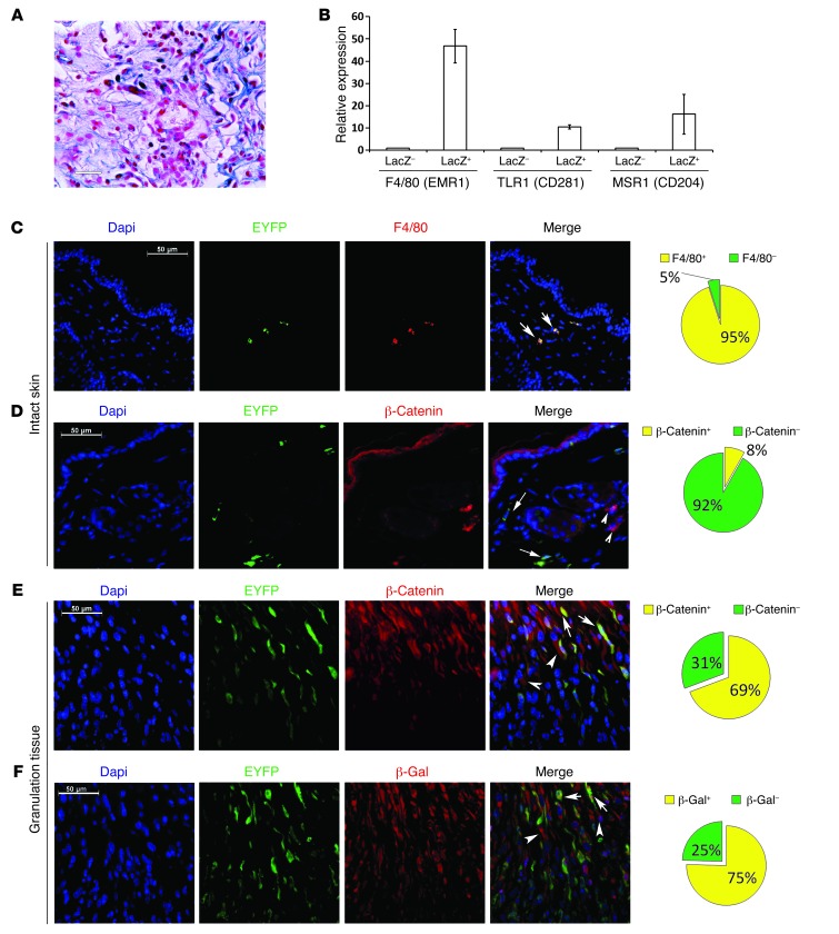
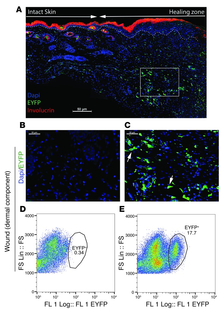
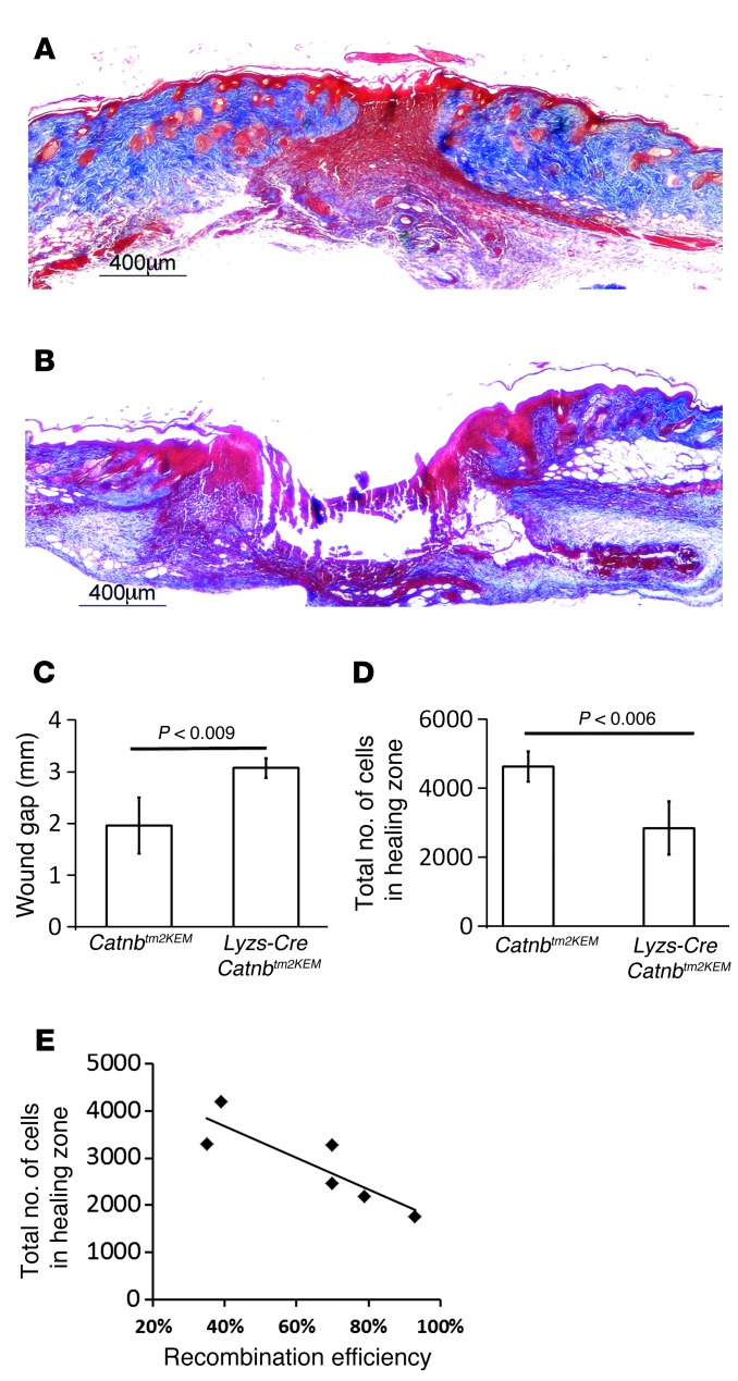
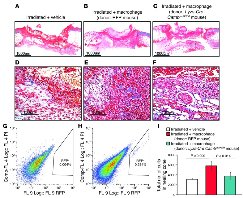
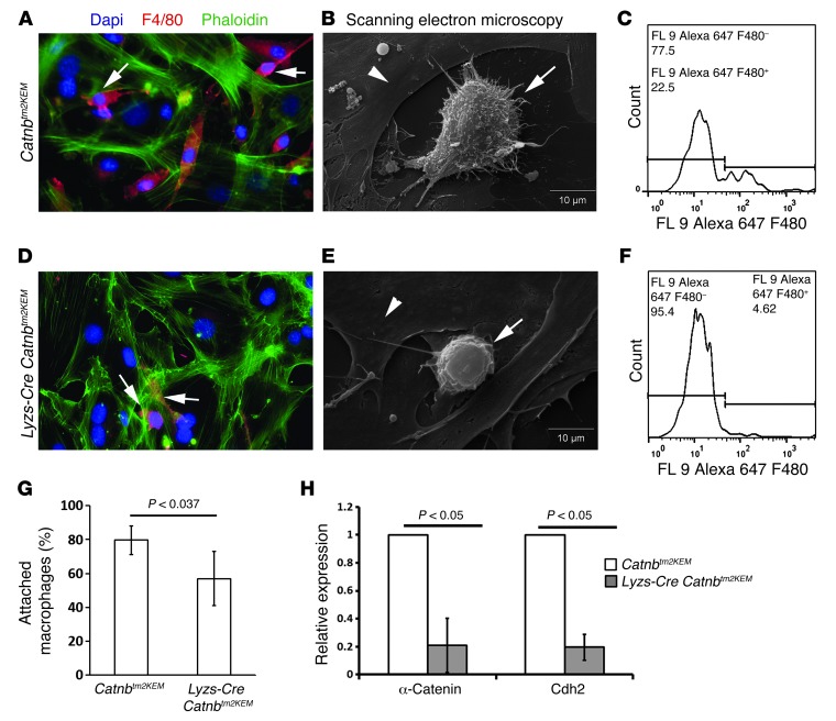
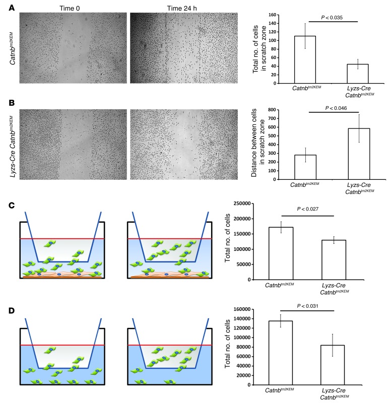
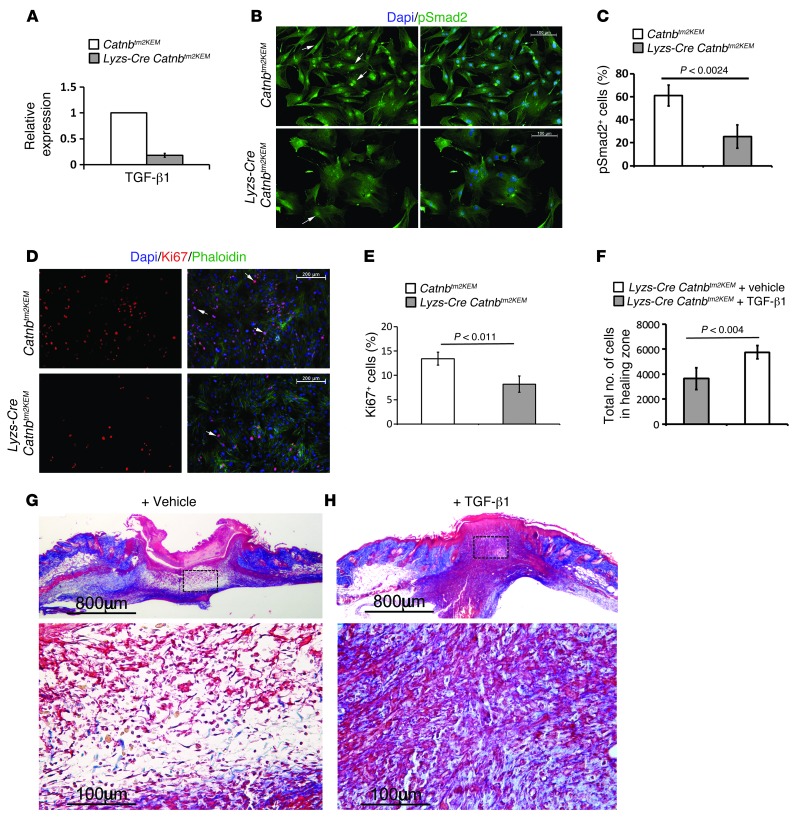
A β-catenin/T cell factor-dependent transcriptional program is critical during cutaneous wound repair for the regulation of scar size; however, the relative contribution of β-catenin activity and function in specific cell types in the granulation tissue during the healing process is unknown. Here, cell lineage tracing revealed that cells in which β-catenin is transcriptionally active express a gene profile that is characteristic of the myeloid lineage. Mice harboring a macrophage-specific deletion of the gene encoding β-catenin exhibited insufficient skin wound healing due to macrophage-specific defects in migration, adhesion to fibroblasts, and ability to produce TGF-β1. In irradiated mice, only macrophages expressing β-catenin were able to rescue wound-healing deficiency. Evaluation of scar tissue collected from patients with hypertrophic and normal scars revealed a correlation between the number of macrophages within the wound, β-catenin levels, and cellularity. Our data indicate that β-catenin regulates myeloid cell motility and adhesion and that β-catenin-mediated macrophage motility contributes to the number of mesenchymal cells and ultimate scar size following cutaneous injury.


Figures

Figure 1
Figure 1. Tcf transcriptionally active cells express genes characteristic of macrophages during skin healing.
(A) Dermal component of a healing wound in a Tcf reporter mouse showing a subpopulation of fibroblast-like cells that were transcriptionally active for β-catenin/Tcf labeled with β-gal. Scale bar: 26 μm. (B) Quantitative RT-PCR analysis showing a higher expression level of genes known to be expressed in macrophages in β-gal–positive cells compared with expression levels of these genes in β-gal–negative cells. Data are shown as the mean ± 95% CI of results from 8 mice. (C) Double immunofluorescence staining of intact skin from a Lysz-Cre ROSA-EYFP mouse showing that EYFP-positive cells were also positive for F4/80. Arrows indicate EYFP-positive myeloid cells. In unwounded mice, EYFP-positive cells were also positive for F4/80. (D) Double immunofluorescence staining of intact skin from a Lysz-Cre ROSA-EYFP mouse showing that macrophages (EYFP-positive cells) in the unwounded skin did not express β-catenin. Arrows show EYFP-positive cells, and arrowheads show EYFP- and β-catenin–positive cells. (E) Double immunofluorescence staining of granulation tissue of healing wounds from a Lysz-Cre ROSA-EYFP mouse showing colocalization of EYFP and β-catenin in EYFP-positive cells. Arrows show EYFP-positive/β-catenin–positive cells, and arrowheads show EYFP-negative/β-catenin–positive cells, indicating that β-catenin was expressed in myeloid cells during the healing process. (F) Double immunofluorescence staining of the wound granulation tissue from a Lysz-Cre ROSA-EYFP Tcf mouse showing colocalization of EYFP and β-gal. Arrows show EYFP-positive/β-gal–positive cells, and arrowheads show EYFP-negative/β-gal–positive cells, indicating that myeloid cells exhibited β-catenin–dependent Tcf-mediated transcriptional activity during healing. (CF) Pie charts illustrating the proportion of positive and negative stained cells in samples from 8 mice. Scale bars: 50 μm.
Figure 2
Figure 2. Lysz-expressing myeloid cells contribute to the dermal compartment of wound repair.
(A) Low-magnification image of the entire wound from a Lysz-Cre ROSA-EYFP mouse showing the granulation tissue and surrounding intact skin. Involucrin (marker of the differentiated upper layer of a keratinocyte) is stained red. Dashed line separates the keratinocyte zone from the lower dermis layer. Arrows separate the intact skin from the healing zone. (B) Fluorescence microscopy of the healing tissue in a ROSA-EYFP control mouse showing the absence of EYFP-positive cells. (C) Fluorescence microscopy of a 7-day-old wound from a Lysz-Cre ROSA-EYFP reporter mouse showing that a subpopulation of cells were EYFP positive. (D) Flow cytometric analysis of the granulation tissue from a control ROSA-EYFP mouse. (E) Flow cytometric analysis of healing tissue in a Lysz-Cre ROSA-EYFP mouse showing that 18% of cells were EYFP positive (Lysz-expressing progeny) in healing skin. (AC) Scale bars: 50 μm. FS, forward scatter; FS Lin: forward scatter on linear scatter; FL1, first fluorescent detector.
Figure 3
Figure 3. Wounds from mice lacking β-catenin in macrophages do not heal.
Trichrome-stained histology of healing wounds. Seven days after wounding, there was a lack of cells in the region that normally contains wound granulation tissue in Lysz-Cre Catnbtm2KEM mice (lacking β-catenin in myeloid cells) compared with that seen in their control littermates. (A) Representative histologic section of a healing wound in a control mouse. Scale bar: 400 μm. (B) Representative histologic section of a healing wound in a mouse in which β-catenin was depleted from macrophages. Scale bar: 400 μm. (C) Wound gap measurements show a larger gap in the Lysz-Cre Catnbtm2KEM mice compared with that seen in control mice. (D) Cell density quantification shows a significant decrease in the number of cells in a healing wound in Lysz-Cre Catnbtm2KEM mice compared with the number of cells observed in control mice. (E) The percentage of bone marrow–derived EYFP-positive macrophages in Lysz-Cre Catnbtm2KEM ROSA-EYFP mice inversely correlated with the total number of cells in the healing wound (correlation coefficient = –0.865, P < 0.014).
Figure 4
Figure 4. Macrophages deficient in β-catenin cannot rescue deficient wound healing in irradiated mice.
Representative histology of healing wounds from an irradiated mouse treated with macrophages from a donor mouse showing partial rescue of the wound phenotype and a significant increase in the number of cells in the wound (AC are low-magnification images; scale bars:1,000 μm. DF are higher-magnification images; scale bars: 100 μm). Macrophages lacking β-catenin were not able to rescue the phenotype. (A and D) Irradiated control mouse treated with carrier only. (B and E) Irradiated mouse treated with macrophages obtained from the bone marrow of a control mouse. (C and F) Irradiated mouse treated with macrophages from Lysz-Cre Catnbtm2KEM mice lacking β-catenin. RFP-labeled macrophages were absent in the healing tissue of control-treated mice (G), while they were present in the healing tissue (H) of the treated mice 1 week after wounding. (I) Quantification of the total number of cells in the healing zone in the various mouse wounds. Data are from 8 mice and are shown as the mean ± 95% CI. Comp-FL, compensated fluorescence.
Figure 5
Figure 5. β-Catenin regulates macrophage cell adhesion to fibroblasts.
Adhesion assay using control macrophages (AC) and macrophages lacking β-catenin (DF). Double immunofluorescence staining of macrophages and fibroblasts in cocultures. More F4/80-positive cells were observed in cocultures with control macrophages (A) in comparison with macrophages lacking β-catenin (D). Arrow shows F4/80-positive cells (red) that remained attached to the fibroblast layer (green). Scanning electron microscopy of macrophages that remained adherent to the fibroblast layer showing that macrophages that lacked β-catenin (E) had less pseudopod-like projections in comparison with control macrophages (B). Arrows show macrophages, and arrowhead shows fibroblasts. Flow cytometric analysis of washed and trypsinized cultured cells showing that there was a greater decrease in the percentage of F4/80-positive macrophages in macrophages lacking β-catenin (F) than in controls (C). (G) Quantification of the number of attached macrophages on fibroblasts, indicating that fewer macrophages lacking β-catenin adhered to fibroblasts. (H) Macrophages lacking β-catenin had lower expression levels of α-catenin and cadherin-2 (N-cadherin) compared with the levels observed in control macrophages. Data represent the mean ± 95% CI for 7 mice.
Figure 6
Figure 6. β-Catenin regulates macrophage migration.
Representative photomicrographs of a scratch assay performed with (A) Lyzs-Cre ROSA-EYFP macrophages and (B) macrophages from Lyzs-Cre Catnbtm2KEM ROSA-EYFP mice showing cell migration immediately after scratching and after 24 hours. Migration was quantified by counting the number of cells migrating into the scratch and by measuring the gap. Bar graphs show the means and 95% CI for the number of cells in the scratch zone and the average distance between the 2 edges of the scratch after 24 hours. (C) Boyden migration assay with macrophages seeded in the upper chamber and fibroblasts in the lower chamber. The total number of cells in the lower chamber was significantly lower when macrophages from Lyzs-Cre Catnbtm2KEM ROSA-EYFP mice were used compared with the total number in control macrophages. (D) Boyden migration assay using macrophages in the upper chamber, with no cells in the lower chamber. The number of migrated cells in the lower chamber was significantly smaller when macrophages from Lyzs-Cre Catnbtm2KEM ROSA-EYFP mice were used compared with the number observed in control macrophages. Graphs show the mean ± 95% CI of data from macrophages from 4 mice in each group.
Figure 7
Figure 7. Macrophages lacking β-catenin induce less TGF-β1 signaling, and TGF-β1 partially rescues the wound phenotype of mice lacking β-catenin in macrophages.
(A) Quantitative RT-PCR analysis showing decreased TGF-β1 expression in β-catenin–deficient macrophages from Lysz-Cre Catnbtm2KEM ROSA-EYFP mice compared with that seen in their control littermates. (B) pSmad2 staining of fibroblasts that were exposed to either control (top panels) or β-catenin–deficient macrophages (bottom panels), indicating less pSmad2-positive cells in fibroblasts that were exposed to macrophages lacking β-catenin (quantified in C). Arrow shows pSmad2-positive cells. Scale bars: 100 μm. (D) Ki67 staining of fibroblasts that were exposed to either control macrophages (top panels) or to β-catenin–deficient macrophages (lower panels), indicating less Ki67-positive cells in fibroblasts that were exposed to macrophages lacking β-catenin (quantified in E). Arrow shows Ki67-positive cells. Scale bars: 200 μm. (F) Cell density quantification shows a significant increase in the number of cells in healing wounds in mice treated with TGF-β1 compared with those treated with vehicle. (G and H) Representative histology of a wound from a mouse treated with vehicle or TGF-β1 whose macrophages lacked β-catenin. Image in G is magnified in the lower panel; scale bars: 800 μm; 100 μm. Image in H is magnified in the lower panel; scale bars: 800 μm; 100 μm. Data represent the mean ± 95% CI of 7 mice.

References

    1. Sen CK, et al. Human skin wounds: a major and snowballing threat to public health and the economy. Wound Repair Regen. 2009;17(6):763–771. doi: 10.1111/j.1524-475X.2009.00543.x. - DOI - PMC - PubMed
    1. Gottrup F. A specialized wound-healing center concept: importance of a multidisciplinary department structure and surgical treatment facilities in the treatment of chronic wounds. Am J Surg. 2004;187(5A):38S–43S. - PubMed
    1. Gurtner GC, Werner S, Barrandon Y, Longaker MT. Wound repair and regeneration. Nature. 2008;453(7193):314–321. doi: 10.1038/nature07039. - DOI - PubMed
    1. Bielefeld KA, Amini-Nik S, Alman BA. Cutaneous wound healing: recruiting developmental pathways for regeneration. Cell Mol Life Sci. 2013;70(12):2059–2081. doi: 10.1007/s00018-012-1152-9. - DOI - PMC - PubMed
    1. Cheon SS, et al. β-Catenin stabilization dysregulates mesenchymal cell proliferation, motility, and invasiveness and causes aggressive fibromatosis and hyperplastic cutaneous wounds. Proc Natl Acad Sci U S A. 2002;99(10):6973–6978. doi: 10.1073/pnas.102657399. - DOI - PMC - PubMed

Publication types

MeSH terms