. 2014 Jun;124(6):2472-82.
doi: 10.1172/JCI69672. Epub 2014 May 16.

Synthetic triterpenoid induces 15-PGDH expression and suppresses inflammation-driven colon carcinogenesis

Synthetic triterpenoid induces 15-PGDH expression and suppresses inflammation-driven colon carcinogenesis

Sung Hee Choi et al. J Clin Invest. 2014 Jun.

Abstract

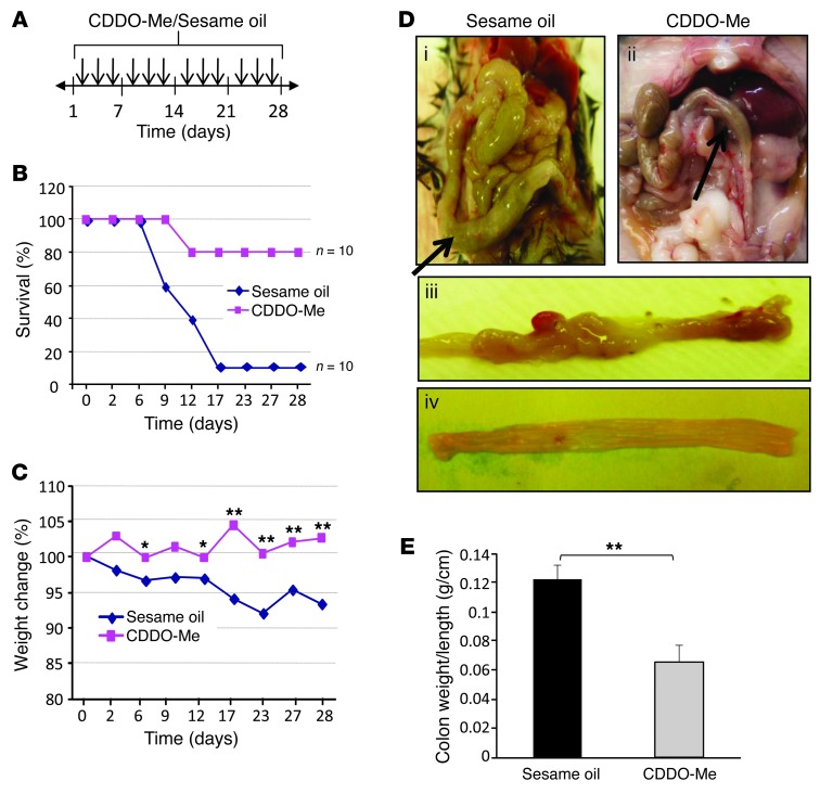
Colitis-associated colon cancer (CAC) develops as a result of inflammation-induced epithelial transformation, which occurs in response to inflammatory cytokine-dependent downregulation of 15-hydroxyprostaglandin dehydrogenase (15-PGDH) and subsequent suppression of prostaglandin metabolism. Agents that both enhance 15-PGDH expression and suppress cyclooxygenase-2 (COX-2) production may more effectively prevent CAC. Synthetic triterpenoids are a class of small molecules that suppress COX-2 as well as inflammatory cytokine signaling. Here, we found that administration of the synthetic triterpenoid 2-cyano-3,12-dioxooleana-1,9(11)-dien-C28-methyl ester (CDDO-Me) suppresses CAC in mice. In a spontaneous, inflammation-driven intestinal neoplasia model, deletion of Smad4 specifically in T cells led to progressive production of inflammatory cytokines, including TNF-α, IFN-γ, iNOS, IL-6, IL-1β; as well as activation of STAT1 and STAT3; along with suppression of 15-PGDH expression. Oral administration of CDDO-Me to mice with SMAD4-deficient T cells increased survival and suppressed intestinal epithelial neoplasia by decreasing production of inflammatory mediators and increasing expression of 15-PGDH. Induction of 15-PGDH by CDDO-Me was dose dependent in epithelial cells and was abrogated following treatment with TGF-β signaling inhibitors in vitro. Furthermore, CDDO-Me-dependent 15-PGDH induction was not observed in Smad3-/- mice. Similarly, CDDO-Me suppressed azoxymethane plus dextran sodium sulfate-induced carcinogenesis in wild-type animals, highlighting the potential of small molecules of the triterpenoid family as effective agents for the chemoprevention of CAC in humans.


Figures

Figure 1
Figure 1. Smad4Tko mice develop CAC.
Biomarkers of disease progression were defined in wild-type and Smad4Tko (KO) mice at 3 and 8 months of age. (A) The colon weight per length of 3-month-old or 8-month-old wild-type and Smad4Tko mice. (B) Expression of proinflammatory cytokines (TNF-α, IL-1β, IL-6, and IFN-γ) from colon epithelial cells from wild-type or KO mice, as measured by RT-PCR. (C) Phosphorylation of STAT1, STAT3, and Iκb as well as expression of iNOS, COX-2, and 15-PGDH, as detected by Western blot. (D) Concentration of nitrate from sera of wild-type and Smad4Tko mice. Analyses at 3 months include wild-type mice (n = 4) and Smad4Tko mice (n = 5); analyses at 8 months include wild-type (n = 7) and Smad4Tko mice (n = 8). Results shown are representative of 4 separate experiments.
Figure 2
Figure 2. Oral administration of CDDO-Me suppresses mucosal inflammation in 8-month-old Smad4Tko mice.
(A) Eight-month-old Smad4Tko mice were given CDDO-Me by gavage every other day for 1 month. (B) Mortality (n = 10 per group). (C) Percent weight change. (D) Colon histology (i and iii show sections from Smad4Tko mice receiving control sesame oil; ii and iv show sections from Smad4Tko mice treated with CDDO-Me). (E) Colon weight per length (g/cm) (n = 9). Error bars indicate SEM; *P < 0.05, **P < 0.01 compared with mice in control group receiving sesame oil alone.
Figure 3
Figure 3. Cancer chemoprevention by CDDO-Me in the Smad4Tko mouse model of CAC.
(A) Mucosal histology was examined in Smad4Tko mice treated with either sesame-oil or CDDO-Me. Paraffin-embedded sections were stained with H&E. Scale bars: 200 μm. (B) Percentage of tumor-bearing mice. (C) Tumor number. (D) Tumor sizes were determined using a digital eyepiece. (E) Histogram showing size distribution of tumors. (F) Expression of proinflammatory mediators in tumor and nontumor intestinal mucosa. Whole tumor specimens and adjacent mucosa samples were homogenized. iNOS, p-STAT3, p-STAT1, COX-2, p-iκB, and β-actin expression were measured by Western blot. N, nontumor intestinal mucosa; T, tumor tissue. ***P < 0.001.
Figure 4
Figure 4. CDDO-Me treatment suppresses inflammatory mediators and increases the expression of 15-PGDH in colon mucosa.
Eight-month-old Smad4Tko mice were treated with either sesame oil or CDDO-Me (250 ng per mouse per day) for 1 month. (A) The percentage of Ki-67–positive cells among all colon epithelium cells was determined in mice treated with either sesame oil or CDDO-Me. Data represent average ± SEM (n = 9). ***P < 0.001 with direct comparison to Smad4Tko mice receiving sesame oil alone. Scale bar: 100 μM. (B) Expression of proinflammatory cytokines, IL-6 and IFN-γ, was measured by RT-PCR. (C) Expression of STAT1 and STAT3, phospho-STAT1, and phospho-STAT3 was measured by Western blot (lanes were run on the same gel but were noncontiguous). (D) Protein and mRNA expression of iNOS and 15-PGDH were measured by Western blot and RT-PCR analysis (lanes were run on the same gel but were noncontiguous). (E) Nitrate concentration in sera of Smad4Tko mice receiving either sesame oil or CDDO-Me. Wild-type control mice also received sesame oil (n = 9).
Figure 5
Figure 5. Chemoprevention of AOM/DSS-induced colon cancer by CDDO-Me.
Body weight and tumor number in mice treated with AOM/DSS or AOM/DSS plus CDDO-Me. (A) Experimental procedure used to induce colon cancer in C57BL/6 mice. After initial AOM injection (10 mg/kg), DSS was given in drinking water, followed by normal drinking water. Either CDDO-Me (250 ng per mouse per day) or sesame oil were given to the mice every other day during the DSS treatment. (B) Changes in body weight among each group were defined as follows: percentage = (final weight – initial weight)/initial weight × 100. CDDO-ME treatment did not affect the body weights. Tumors were enumerated examined using a dissecting microscope at day 53. (C) Images of colons at necropsy and (D) H&E staining of tumor morphology. Scale bar: 200 μM. (E) Colon length and (F) number of tumors in sesame oil–treated or CDDO-ME–treated groups. The sesame oil–treated group had 10 mice, and the CDDO-ME–treated group had 9 mice. Results are mean ± SEM. **P < 0.01.
Figure 6
Figure 6. CDDO-Me increases the expression of 15-PGDH in colon epithelial cells.
(A) Expression of 15-PGDH protein and mRNA after treatment with CDDO-Me. FET cells were treated with various doses of CDDO-Me (0–300 nM) for 24 hours. Expression of 15-PGDH protein and mRNA was analyzed by Western blotting and RT-PCR analysis (β-actin was used as a control). Lanes were run on the same gel but were noncontiguous. The data shown are representative of 6 independent experiments. (B) Time-dependent effect of CDDO-Me (100 nM) on 15-PGDH expression. FET cells were treated with CDDO-Me (100 nM) for 6, 12, 24, 48, and 72 hours and analyzed by Western blot. The data shown are representative of 3 independent experiments. (C) CDDO-Me–induced 15-PGDH promoter luciferase activity. pGL3 and 15-PGDH promoter activity in FET cells following stimulation with CDDO-Me (300 nM) for 24 hours. The fold induction of the relative levels of 15-PGDH transcripts was compared with that of untreated pGL3 transcripts. A dual luciferase assay was performed, and data shown are averages of triplicate independent measurements of Firefly/Renilla luciferase readings normalized to untreated controls. Data represent average ± SEM (n = 4–6). (D) CDDO-Me inhibited colon epithelial cellular proliferation in a dose-dependent manner. 5 × 10³ FET cells were cultured with CDDO-Me (0–300 nM) in 96-well plates with treatment, and proliferation was assessed by incorporation of 3H-thymidine.
Figure 7
Figure 7. Induction of 15-PGDH expression by CDDO-Me requires SMAD-dependent TGF-β signaling.
(A) FET cells cultured with CDDO-Me plus TGF-β (1 ng/ml). Western blot is representative of 3 independent experiments (noncontiguous lanes were run on the same gel). Graphs represent the mean ± SEM of 4 independent sets of experiments. (B) FET cells transfected with SBE-luc were treated with CDDO-Me plus TGF-β (1 ng/ml). Results of a dual luciferase assay are shown as averages (triplicate independent measurements of Firefly/Renilla luciferase normalized to untreated controls). Results are representative of 3 different experiments. (C) FET cells were treated with either TGF-β or CDDO-Me (for 10 minutes to 9 hours) and phosphorylation of SMAD2 and SMAD3 was examined by Western blot. (D) Cells were incubated for 30 minutes with either TGF-β receptor inhibitors, SB431542 (10 μM) or IN1130 (10 μM), or SMAD3-specific inhibitor, SIS3 (10 μM), before adding TGF-β (1 ng/ml) and/or CDDO-Me (300 nM). Data represent 3 independent experiments (all noncontiguous lanes were run on the same gel). (E) CDDO-Me failed to induce mucosal 15-PGDH expression in vivo in SMAD3 KO mice. Mice received CDDO-Me (1.25 μg or 5 μg) by gavage, and colon epithelial scrapings were analyzed by Western blot 24 hours after the last dose (all noncontiguous lanes were run on the same gel). (F) Proliferation of FET cells (with or without CDDO-Me and/or TGF-β) was measured by incorporation of 3H-thymidine.

Comment in

Similar articles

Cited by

References

    1. Kappelman MD, Moore KR, Allen JK, Cook SF. Recent trends in the prevalence of Crohn’s disease and ulcerative colitis in a commercially insured US population. Dig Dis Sci. 2012;58(2):519–525. - PMC - PubMed
    1. [No authors listed] Epidemiological and clinical features of Spanish patients with Crohn’s disease. Spanish Epidemiological and Economic Study Group on Crohn’s disease. Eur J Gastroenterol Hepatol. 1999;11(10):1121–1127. doi: 10.1097/00042737-199910000-00008. - DOI - PubMed
    1. Molodecky NA, et al. Increasing incidence and prevalence of the inflammatory bowel diseases with time, based on systematic review. Gastroenterology. 2012;142(1):46–54. doi: 10.1053/j.gastro.2011.10.001. - DOI - PubMed
    1. Gersemann M, Wehkamp J, Stange EF. Innate immune dysfunction in inflammatory bowel disease. J Intern Med. 2012;271(5):421–428. doi: 10.1111/j.1365-2796.2012.02515.x. - DOI - PubMed
    1. Boirivant M, Cossu A. Inflammatory bowel disease. Oral Dis. 2012;18(1):1–15. doi: 10.1111/j.1601-0825.2011.01811.x. - DOI - PubMed

Publication types

MeSH terms