. 2014 Jun;124(6):2750-61.
doi: 10.1172/JCI74604. Epub 2014 May 16.

Elevated sphingosine-1-phosphate promotes sickling and sickle cell disease progression

Elevated sphingosine-1-phosphate promotes sickling and sickle cell disease progression

Yujin Zhang et al. J Clin Invest. 2014 Jun.

Abstract

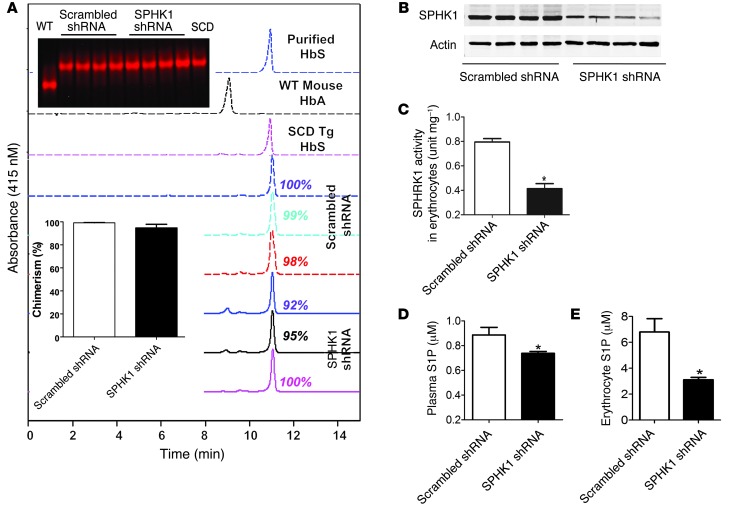
Sphingosine-1-phosphate (S1P) is a bioactive lipid that regulates multicellular functions through interactions with its receptors on cell surfaces. S1P is enriched and stored in erythrocytes; however, it is not clear whether alterations in S1P are involved in the prevalent and debilitating hemolytic disorder sickle cell disease (SCD). Here, using metabolomic screening, we found that S1P is highly elevated in the blood of mice and humans with SCD. In murine models of SCD, we demonstrated that elevated erythrocyte sphingosine kinase 1 (SPHK1) underlies sickling and disease progression by increasing S1P levels in the blood. Additionally, we observed elevated SPHK1 activity in erythrocytes and increased S1P in blood collected from patients with SCD and demonstrated a direct impact of elevated SPHK1-mediated production of S1P on sickling that was independent of S1P receptor activation in isolated erythrocytes. Together, our findings provide insights into erythrocyte pathophysiology, revealing that a SPHK1-mediated elevation of S1P contributes to sickling and promotes disease progression, and highlight potential therapeutic opportunities for SCD.


Figures

Figure 1
Figure 1. Metabolomic screening reveals that blood S1P levels and erythrocyte SPHK1 activity are elevated in SCD Tg mice.
(A) Z-score quantification of lipids detected in the blood of both WT and SCD Tg mice. Among all lipids detected, S1P was one of the most substantially elevated in the blood of SCD Tg mice compared with that in WT mice (n = 6). (B and C) LC/MS POS platform measurement indicates that S1P levels in the (B) erythrocytes and (C) plasma of SCD mice were significantly elevated compared with those in WT mice (n = 6–8). (D) Total erythrocyte SPHK1 activity was significantly elevated in SCD Tg mice compared with that in WT mice (n = 8). (E) SPHK1 activities in purified reticulocytes and mature erythrocytes of SCD Tg mice. Values shown represent the mean ± SEM (n = 6–8). *P < 0.05 versus WT.
Figure 2
Figure 2. PF-543, a potent, specific SPHK1 inhibitor, reduces sickling, hemolysis, and inflammation in SCD Tg mice by reducing erythrocyte SPHK1 activity and S1P levels.
(AC) PF-543 treatment significantly reduced (A) erythrocyte SPHK1 activity, (B) erythrocyte levels, and (C) plasma levels of S1P in SCD Tg mice. (D) Representative blood smears of SCD Tg mice, as a function PF-543 treatment (original magnification, ×100). (E) Percentages of sickle cells and reticulocytes were significantly reduced by PF-543 treatment in SCD Tg mice. (FI) Effects of PF-543 treatment on (F) plasma Hb, (G) plasma haptoglobin, (H) plasma total bilirubin, and (I) circulating cytokines in SCD Tg mice. Values shown represent the mean ± SEM (n = 6–11). *P < 0.05 versus SCD Tg mice treated with DMSO.
Figure 3
Figure 3. Specific knockdown of SPHK1 in HSCs in BMT SCD chimeras reduced erythrocyte SPHK1 protein levels and erythrocyte and plasma S1P levels.
(A) HPLC and electrophoresis analysis of HbS and mouse normal Hb (HbA) in BMT SCD chimeras to assess the percentage of chimerism. Representative electrophoresis analysis and the average of percentage of chimerism with HbS in SCD chimeras were shown as insets. Data shown represent the mean ± SEM (n = 6–9). (B) Erythrocyte SPHK1 levels, (C) activity, (D) plasma S1P levels, and (E) erythrocyte S1P levels were significantly reduced in the SCD chimeras with specific SPHK1 knockdown compared with those in mice with BMCs infected with a scrambled shRNA. *P < 0.05 versus SCD chimeras with BMCs infected with recombinant lentivirus encoding scrambled shrna.
Figure 4
Figure 4. Specific knockdown of SPHK1 in HSCs in BMT SCD chimeras reduced sickling, hemolysis, inflammation, and prolonged life span of erythrocytes.
(A) Blood smears of SCD chimeras with or without SPHK1 knockdown (original magnification, ×100). (B) Percentages of sickle cells and reticulocytes were significantly reduced in the SCD chimeras with HSC-specific SPHK1 knockdown. (CE) SPHK1 knockdown in HSCs (C) decreased plasma Hb levels, (D) prolonged life span of erythrocytes, and (E) reduced circulating cytokines in SCD chimeras. Values shown represent the mean ± SEM (n = 6–11). *P < 0.05 versus SCD chimeras with BMCs infected with recombinant lentivirus encoding scrambled shrna.
Figure 5
Figure 5. Specific knockdown of SPHK1 in HSCs in BMT SCD chimeras reduced splenomegaly and tissue damage.
(A) Spleen size and (B) H&E staining of spleens, livers, and lungs of SCD chimeras with HSC-specific SPHK1 knockdown and controls. (C) Representative Evans blue staining and (D) quantification of its concentration in the lungs of the control SCD chimeras and SCD chimeras with specific SPHK1 knockdown. (E) Albumin concentrations in bronchial alveolar lavage (BAL) fluid collected from the control SCD chimeras and SCD chimeras with specific SPHK1 knockdown. Values shown represent the mean ± SEM (n = 6–11). *P < 0.05 versus SCD chimeras with BMCs infected with recombinant lentivirus encoding scrambled shrna. Scale bar: 200 μM.
Figure 6
Figure 6. Specific knockdown of SPHK1 in HSCs in BMT SCD chimeras increased survival rates and reduced pulmonary congestion and inflammation under hypoxic conditions.
(A) Knockdown of SPHK1 in HSCs prolonged survival rates of BMT SCD chimeras under hypoxic conditions with 8% O2 concentration. (B and C) Knockdown of SPHK1 in HSCs in SCD chimeras (B) reduced hypoxia-induced pulmonary congestion and (C) decreased elevation of multiple cytokines in the lung tissue. Values shown represent the mean ± SEM (n = 6–9). *P < 0.05 versus SCD chimeras with BMCs infected with recombinant lentivirus encoding scrambled shRNA. Scale bar: 200 μM.
Figure 7
Figure 7. Erythrocyte SPHK1 activity and S1P levels in both erythrocytes and plasma are elevated in individuals with SCD.
(A and B) Average S1P levels in (A) erythrocytes and (B) plasma from healthy volunteers (control, n = 14) and patients with SCD (n = 30). (C) Erythrocyte SPHK1 activity for healthy volunteers (control, n = 14) and patients with SCD (n = 30). Data are presented as the mean ± SEM. *P < 0.05 relative to the control individuals.
Figure 8
Figure 8. SPHK1-mediated elevation of S1P contributes directly to hypoxia-induced sickling in cultured human sickle erythrocytes independent of S1P receptor activation.
(AC) Pretreatment of cultured primary erythrocytes isolated from patients with SCD with PF-543 inhibited hypoxia-mediated induction of (A) SPHK1 activity, (B) S1P production, and (C) sickling in a dosage-dependent manner. (D) Changes in the percentage of sickled cells in erythrocytes isolated from patients with SCD, following exposure to different hypoxic conditions in the absence or presence of PF-543 treatment. (E) S1P contributes to sickling independent of S1P receptor activation. S1P receptor antagonists (VPC, S1P1 and S1P3 receptor antagonist; JTE, S1P2 receptor antagonist) did not reduce hypoxia-induced sickling. (F) Exogenous S1P (100 to 500 nM) did not enhance hypoxia-induced sickling in cultured human sickle erythrocytes under different hypoxia conditions. Data are presented as the mean ± SEM. *P < 0.05 relative to the samples under normoxic conditions, **P < 0.05 relative to untreated hypoxia samples, #P < 0.05 relative to PF-543–treated samples at lower concentration (n = 5–6). (G) Under hypoxic conditions, increased S1P, due to elevated erythrocyte SPHK1 activity, contributes to sickling independent of its receptor activation. Because erythrocytes store the highest amount of S1P (8, 9), the hemolysis associated with sickling unleashes massive amounts of this pleiotropic signaling molecule with widespread detrimental effects, including inflammation and tissue injury at multiple sites. Without interference, increased sickling, hemolysis, inflammation, and tissue injury function as a malicious cycle, leading to more severe hypoxia and further elevation of erythrocyte SPHK1 activity and S1P levels. The use of SPHK1 inhibitors to lower S1P levels reduces sickling and represents a potentially important novel mechanism-based therapy for SCD.

References

    1. Madigan C, Malik P. Pathophysiology and therapy for haemoglobinopathies. Part I: sickle cell disease. Expert Rev Mol Med. 2006;8(9):1–23. - PubMed
    1. Urbinati F, Madigan C, Malik P. Pathophysiology and therapy for haemoglobinopathies. Part II: thalassaemias. Expert Rev Mol Med. 2006;8(10):1–26. - PubMed
    1. Ingram VM. Sickle-cell anemia hemoglobin: the molecular biology of the first “molecular disease” — the crucial importance of serendipity. Genetics. 2004;167(1):1–7. doi: 10.1534/genetics.167.1.1. - DOI - PMC - PubMed
    1. Strasser BJ. Perspectives: molecular medicine. “Sickle cell anemia, a molecular disease”. Science. 1999;286(5444):1488–1490. doi: 10.1126/science.286.5444.1488. - DOI - PubMed
    1. Steinberg MH. Sickle cell anemia, the first molecular disease: overview of molecular etiology, pathophysiology, and therapeutic approaches. ScientificWorldJournal. 2008;8:1295–1324. doi: 10.1100/tsw.2008.157. - DOI - PMC - PubMed

Publication types

MeSH terms