. 2014 Jun 19;54(6):1022-1033.
doi: 10.1016/j.molcel.2014.04.011. Epub 2014 May 15.

Catalytic and noncatalytic roles of the CtIP endonuclease in double-strand break end resection

Affiliations

Catalytic and noncatalytic roles of the CtIP endonuclease in double-strand break end resection

Nodar Makharashvili et al. Mol Cell. .

Abstract

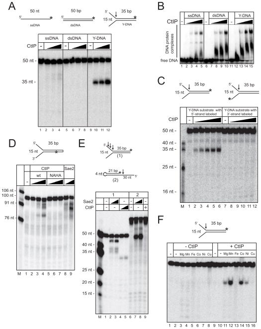
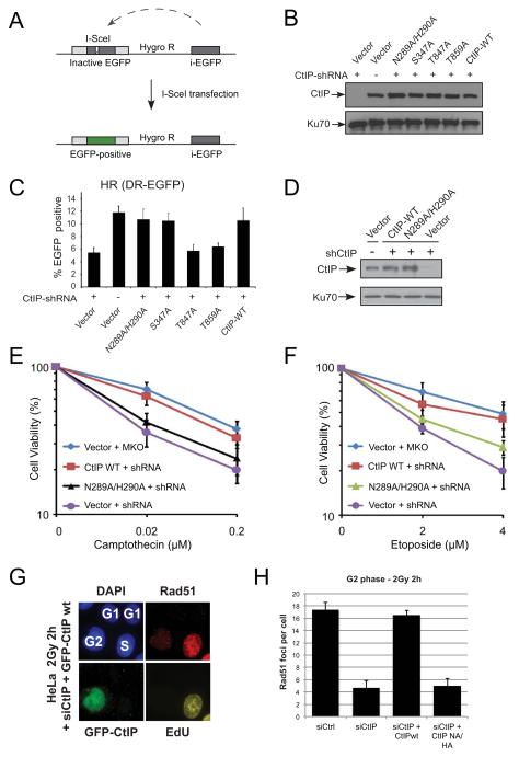
The carboxy-terminal binding protein (CtBP)-interacting protein (CtIP) is known to function in 5' strand resection during homologous recombination, similar to the budding yeast Sae2 protein, but its role in this process is unclear. Here, we characterize recombinant human CtIP and find that it exhibits 5' flap endonuclease activity on branched DNA structures, independent of the MRN complex. Phosphorylation of CtIP at known damage-dependent sites and other sites is essential for its catalytic activity, although the S327 and T847 phosphorylation sites are dispensable. A catalytic mutant of CtIP that is deficient in endonuclease activity exhibits wild-type levels of homologous recombination at restriction enzyme-generated breaks but is deficient in processing topoisomerase adducts and radiation-induced breaks in human cells, suggesting that the nuclease activity of CtIP is specifically required for the removal of DNA adducts at sites of DNA breaks.


Figures

Figure 1
Figure 1. CtIP is a 5′ strand flap endonuclease
(A) Structures of DNA substrates (top): ssDNA, and dsDNA, Y-structure with 15 nt branch. Each substrate contains the same top strand, labeled with [32P] at the 3′ end (asterisk). Nuclease assays were performed with human wt CtIP (50, 100, and 200 nM) in 5 mM MgCl2, and products were separated by denaturing polyacrylamide gel electrophoresis (PAGE). The arrow shows the position of CtIP-mediated cleavage. (B) Gel mobility shift assays were performed with DNA substrates as in (A) with human CtIP (25, 50, 100, or 200 nM), separated by native PAGE. (C) Nuclease assays as in (A) using Y structures labeled on the 3′ end of the top strand or the bottom strand as shown. (D) Nuclease assays with wt CtIP (50, 100, or 200 nM) or wt recombinant Sae2 (1.5 or 6 nM) as in (A) with 5 mM MgCl2 and a Y-structure DNA containing a hairpin and an internal [32P] label as indicated. (E) Nuclease assays with wt CtIP (100 or 200 nM) or wt recombinant Sae2 (3 or 6 nM) in 5 mM MgCl2 and a Y-structure DNA (1) or a hairpin with a 5′ overhang (2). (F) Nuclease assays with wt CtIP (200 nM) on Y-structure DNA as in (A) but with various metals (6 mM Mg2+ or 2 mM other metals, and 1 mM EDTA) as indicated.
Figure 2
Figure 2. Regulation of CtIP nuclease activity by conserved amino acids and phosphorylation
(A) Schematic diagram of the CtIP protein showing known features and phosphorylation sites relevant to this study: S231, S276, T315, and S347 (see Table S1). (B) Nuclease assays with Y-structure as in Fig. 1A, using wt, NA/HA, or H290N CtIP proteins at 25, 50, 100, or 200 nM. (C) Gel mobility shift assays with a 249 bp dsDNA substrate internally labeled with [32P], using wt or NA/HA CtIP at 6.25, 12.5, 25, 50, 100, or 200 nM. Reactions were separated by native PAGE. (D) Quantitation of CtIP endonuclease assays performed as in (B) with 200 nM wt or NA/HA proteins in 5 mM MgCl2 or 1 mM MnCl2 as indicated. The average of 3 experiments is shown with error bars indicating standard deviation. (E, F) Nuclease assays with a Y-structure as in Fig. 1A, using wt and mutant proteins at 100 or 200 nM. (G) Nuclease assays with a Y-structure as in Fig. 1A, using wt and mutant proteins at 50, 100 or 200 nM.
Figure 3
Figure 3. CtIP and Mre11 have N-terminal oxidation-sensitive metal binding sites
(A) Schematic diagram of CtIP showing known features as in Fig. 2 but also indicating regions of recognition by Flag and CtIP antibodies and domain where oxidation takes place (see main text). (B) Human CtIP was incubated with ascorbic acid and H2O2; reactions were stopped at the indicated time points, separated by SDS-PAGE, and either stained with coomassie blue or transferred to a membrane and blotted with anti-Flag or anti-CtIP antibodies as indicated. Red arrows indicate predominant cleavage products. (C) Schematic diagram of Mre11 showing known features and indicating the amino acids that directly contact metal ions in the crystal structure of human Mre11 (Park et al., 2011). (D) Full-length human Mre11 was incubated with ascorbic acid and H2O2; reactions were separated by SDS-PAGE and stained with coomassie blue or probed for the C-terminal Flag epitope as indicated. Red arrows indicate cleavage products with the bold arrow indicating the predominant product. (E) Truncated Mre11 (a.a. 1 to 616) was treated as in (D). Bands marked with black or yellow triangles were identified by N-terminal sequencing as C-terminal fragments starting with a.a. 132 and a.a. 218, respectively (see Table S2).
Figure 4
Figure 4. CtIP nuclease activity is required for DNA end processing and damage survival
(A) Schematic representation of an eGFP-based restriction enzyme induced HR repair reporter assay in U2OS cells (Wang et al., 2012). Expression of I-SceI endonuclease induces a DSB; repair via HR results in expression of green fluorescent eGFP protein. (B) Expression of wt, NA/HA, S347A, T847A, or T859A proteins in U2OS cells with CtIP depleted was analyzed by western blot for CtIP protein, using Ku70 for normalization. (C) U2OS cells from (B) carrying the HR reporter and expressing HA-CtIP wt or indicated mutants, with CtIP depleted by shRNA, were induced with I-SceI and assayed for EGFP-positive events. Error bars indicate standard error from at least 3 independent experiments. (D) Expression of wt or NA/HA proteins in U2OS cells with CtIP depleted was analyzed by western blot for CtIP, using Ku70 for normalization. U2OS cells from (D) were analyzed for cell survival after Camptothecin (CPT) (E) or etoposide (F) mediated DNA damage in U2OS cells expressing HA-CtIP WT or indicated mutants, with CtIP shRNA knockdown or mock control (MKO). (G) Endogenous CtIP was depleted in HeLa cells by siRNA and cells were transfected with GFP-tagged wt or NA/HA CtIP plasmids 48 h before 2 Gy irradiation. Cells were incubated with EdU 30 min prior to IR and during the entire repair period. All EdU-positive S-phase cells were excluded from the analysis and only EdU-negative G2-irradiated cells were analyzed. (H) Rad51 foci were enumerated in GFP-positive G2 cells at 2 h after 2 Gy. Error bars indicate standard error from 3 independent experiments. Also see Fig. S3.

References

    1. Alagoz M, Chiang SC, Sharma A, El-Khamisy SF. ATM deficiency results in accumulation of DNA-topoisomerase I covalent intermediates in neural cells. PloS one. 2013;8:e58239. - PMC - PubMed
    1. Andberg M, Jantti J, Heilimo S, Pihkala P, Paananen A, Koskinen AM, Soderlund H, Linder MB. Cleavage of recombinant proteins at poly-His sequences by Co(II) and Cu(II) Protein science: a publication of the Protein Society. 2007;16:1751–1761. - PMC - PubMed
    1. Arthur LM, Gustausson K, Hopfner KP, Carson CT, Stracker TH, Karcher A, Felton D, Weitzman MD, Tainer J, Carney JP. Structural and functional analysis of Mre11–3. Nucleic acids research. 2004;32:1886–1893. - PMC - PubMed
    1. Beucher A, Birraux J, Tchouandong L, Barton O, Shibata A, Conrad S, Goodarzi AA, Krempler A, Jeggo PA, Lobrich M. ATM and Artemis promote homologous recombination of radiation-induced DNA double-strand breaks in G2. EMBO J. 2009;28:3413–3427. - PMC - PubMed
    1. Cejka P, Cannavo E, Polaczek P, Masuda-Sasa T, Pokharel S, Campbell JL, Kowalczykowski SC. DNA end resection by Dna2-Sgs1-RPA and its stimulation by Top3-Rmi1 and Mre11-Rad50-Xrs2. Nature. 2010;467:112–116. - PMC - PubMed

Publication types

MeSH terms

LinkOut - more resources