. 2014 Jul 11;289(28):19383-94.
doi: 10.1074/jbc.M114.575159. Epub 2014 May 16.

Calpain 5 is highly expressed in the central nervous system (CNS), carries dual nuclear localization signals, and is associated with nuclear promyelocytic leukemia protein bodies

Affiliations

Calpain 5 is highly expressed in the central nervous system (CNS), carries dual nuclear localization signals, and is associated with nuclear promyelocytic leukemia protein bodies

Ranjana Singh et al. J Biol Chem. .

Abstract

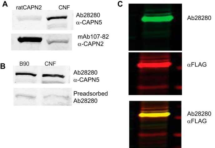
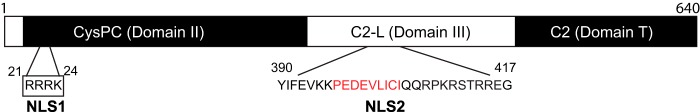
Calpain 5 (CAPN5) is a non-classical member of the calpain family. It lacks the EF hand motif characteristic of classical calpains but retains catalytic and Ca(2+) binding domains, and it contains a unique C-terminal domain. TRA-3, an ortholog of CAPN5, has been shown to be involved in necrotic cell death in Caenorhabditis elegans. CAPN5 is expressed throughout the CNS, but its expression relative to other calpains and subcellular distribution has not been investigated previously. Based on relative mRNA levels, Capn5 is the second most highly expressed calpain in the rat CNS, with Capn2 mRNA being the most abundant. Unlike classical calpains, CAPN5 is a non-cytosolic protein localized to the nucleus and extra-nuclear locations. CAPN5 possesses two nuclear localization signals (NLS): an N-terminal monopartite NLS and a unique bipartite NLS closer to the C terminus. The C-terminal NLS contains a SUMO-interacting motif that contributes to nuclear localization, and mutation or deletion of both NLS renders CAPN5 exclusively cytosolic. Dual NLS motifs are common among transcription factors. Interestingly, CAPN5 is found in punctate domains associated with promyelocytic leukemia (PML) protein within the nucleus. PML nuclear bodies are implicated in transcriptional regulation, cell differentiation, cellular response to stress, viral defense, apoptosis, and cell senescence as well as protein sequestration, modification, and degradation. The roles of nuclear CAPN5 remain to be determined.

Keywords: Calpain; Nuclear Transport; Protease; SUMO-interacting Motif (SIM); SUMOylation.


Figures

FIGURE 1.
FIGURE 1.
Calpain 5 is highly expressed calpain in rat brain. Relative mRNA expression of calpains 1, 2, 5, 7, and 10 was calculated using the comparative CT (ΔΔCT) method in 3-month-old male Sprague-Dawley rat brain homogenate (n = 4). ΔCT of each of the calpain isoforms was obtained as a difference in the CT value from an endogenous control, Gapdh. ΔΔCT of the target gene was calculated by subtracting ΔCT of the target gene from the ΔCT value of the reference gene, Capn1. Relative mRNA expression of the target gene was then reported as 2−ΔΔCT. The results show that after Capn2, Capn5 is the highest expressing calpain in the brain followed by calpains 7, 10, and 1. The results are expressed as the group means ± S.D., n = 4.
FIGURE 2.
FIGURE 2.
Ab28280 is specific to calpain 5. A, in the crude nuclear fraction (CNF) from rat brain, Ab28280 detects a band of ∼75 kDa but does not detect purified rat calpain 2. B, anti-calpain 2 (mAb 107-82) detected purified calpain 2 and weak calpain 2 immunoreactivity in the crude nuclear fraction. Preincubation of the Ab28280 antibody with the N-terminal peptide used as the immunogen (Abcam Ab41310) abolished the 75-kDa band immunoreactivity on Western blots of brain homogenate from 90-day-old rats. C, as a positive control, we examined extracts of SH-SY5Y cells expressing full-length human CAPN5 fused with a FLAG tag on the C terminus (p3X-CMVTM-14-hCAPN51–640-FLAG). Cell lysate was prepared 24 h post-transfection and probed through Western blotting. Ab28280, with secondary antibody IRDye 800CW anti-rabbit IgG, detected a similar prominent band of ∼75 kDa using the 800 nm channel of the Li-Cor Oydessy imaging system and is presented as a green band in the Li-Cor system. FLAG, detected with an anti-FLAG-M2 antibody and Alexa Fluor-680 anti-mouse IgG, using the 700 nm channel of the Li-Cor Odyssey, is shown in red. The overlap of the bands detected by the anti-FLAG and Ab28280 (yellow) illustrates detection of recombinant human CAPN5 by Ab28280.
FIGURE 3.
FIGURE 3.
Calpain 5 is present in the nuclei of neurons and glia. A, X-Gal staining was performed on 40-μm coronal brain sections of ∼3-month-old male Capn5+/LacZ mice (n = 6). In the hippocampal formation, X-Gal staining was prominent in all neurons including CA1 and CA3 pyramidal neurons, granule cells of the dentate gyrus (DG), and hilar neurons (DH). X-Gal staining was also present in molecular layers, suggesting expression in glial cells and consistent with the ubiquitous expression of Capn5 mRNA. B–D, immunohistochemical localization of CAPN5. Confocal images were obtained following double immunolabeling of 40-μm coronal ∼3-month-old male Sprague-Dawley rat brain sections. The co-localization of CAPN5 and NeuN (B), a neuronal nuclear protein (82), indicates that CAPN5 is predominantly localized to neuronal nuclei, although faint extranuclear staining was also observed. CAPN5 immunoreactivity was evident in the nuclei of cells positive for GFAP (C), an intermediate filament protein in astrocytes (83). In cells positive for the APC protein (D), a marker for mature oligodendrocytes (84), CAPN5 immunoreactivity was also localized to the nucleus.
FIGURE 4.
FIGURE 4.
Calpain 5 is enriched in the nuclear nucleic acid binding fraction. A, following differential centrifugation, CAPN5 immunoreactivity was prominent in the crude nuclear fraction (CNF) and was not detected in the cytosolic fraction (C). Marker proteins included histone H3 (nuclear) and β-tubulin as a cytosolic marker. B, using the Qiagen nuclear protein kit, rat brain cortex was subfractionated into the cytosolic fraction, the NABP fraction, and the INP fraction followed by probing for CAPN5 and marker proteins by Western blot. CAPN5 was enriched in the NABP fraction but also was detected in the INP fraction. Survival of motor neuron (SMN) protein resides both in the nucleus and the cytosol (85, 86) and was used as a marker for the NABP fraction. Histone H3 is a marker for INP fraction. C, quantitation of the relative intensity of the CAPN5 immunoreactive band in B is shown. The results, reported as group means ± S.D., n = 3, were analyzed as one-way analysis of variance followed by Tukey's multiple comparison test, **, p < 0.001. D, confocal images of 40-μm male Sprague-Dawley rat brain sections co-immunostained with antibodies against CAPN5, NeuN, GFAP, and Hoechst (a nuclear marker) show punctate nuclear localization of CAPN5 in each of the cell types.
FIGURE 5.
FIGURE 5.
Calpain 5 is associated with PML bodies. A, co-immunolabeling of endogenous CAPN5 and PML in SH-SY5Y cells indicates that CAPN5 is localized with PML bodies. PML-independent CAPN5 localization was also observed. With every PML nuclear body, a CAPN5 immunoreactive dot or larger cluster was either co-localized with PML or in very close proximity to PML, as indicated by arrows. However, the size of the PML nuclear body did not appear to correlate with the size of the CAPN5 cluster. Extranuclear CAPN5 immunoreactivity was also observed, particularly in the perinuclear region, indicated by an arrowhead. B, PML nuclear bodies were also associated with recombinant hCAPN5. SH-SY5Y cells were transiently transfected with full-length human CAPN5 cDNA with a C-terminal ZsGreen1 tag (pN1-CAPN51–640-ZsGreen1) using Lipofectamine 2000. At 24 h post-transfection, CAPN5-ZsGreen1 expression was detected in intranuclear punctate domains and as extranuclear aggregates. Immunocytochemistry with α-PML antibody reveals co-localization of CAPN5 clusters with PML nuclear bodies.
FIGURE 6.
FIGURE 6.
RRRK (aa 21–24) in human CAPN5 is a nuclear localization signal (NLS1). SH-SY5Y cells were transiently transfected with the first 30 amino acids of CAPN5 fused to ZsGreen1 or CAPN51–30-ZsGreen1 or with full-length CAPN5-ZsGreen1. A, schematic showing the labels of the various constructs and the location of mutated NLS. B, at 24 h post-transfection, CAPN51–30 was enriched in the nucleus. C, mutation of amino acids 21–24, RRRK, to AAAA shifted enrichment to the cytosol. D and E, full-length CAPN5 appears as dot-like domains in the nucleus (D), with large perinuclear aggregates also observed in many cells (E). F, residues 21–24 in the full-length CAPN5 reduced the number of cells with punctate nuclear bodies and the number of nuclear bodies in some cells. G, other cells showed only cytosolic aggregates.
FIGURE 7.
FIGURE 7.
Calpain 5 NLS2 (aa 390–417). A, transient transfection of SH-SY-5Y cells with pN1-CAPN5390–417-ZsGreen1 (YIFEVKKPEDEVLICIQQRPKRSTRREG-ZsGreen1), a peptide sequence enclosing putative bipartite NLS and upstream residues YIFEV, resulted in the punctate nuclear expression of the fusion protein. B, similar results were obtained with a C-terminal mCherry tag. C, reducing this sequence by one amino acid (pN1-CAPN5391–417-ZsGreen1) abolished the nuclear, but not perinuclear, localization. D, a slightly longer sequence, 388–417, exhibited similar punctate nuclear localization to aa 390–417 (not shown). Mutagenesis of the basic amino acid residues to alanines (PQYIFEVAAPEDEVLICIQQAPAASTAAEG) did not prevent the nuclear localization, although the organization appeared less punctate. E, mutating Ile403 to Ala in the predicted SUMO-interacting motif, PEDEVLICI (YIFEVKKPEDEVLACIQQRPKRSTRREG), aborts the nuclear localization. F, deletion of NLS2 (pN1-CAPN5Δ390–417-mCherry) resulted in diffuse cellular localization in many cells, whereas nuclear localization was observed in other cells. Similar results were observed with a ZsGreen1 C-terminal tag (not shown). G, mutation of NLS1 and deletion of NLS2 (pN1-CPAN521–24AΔ390–417-ZsGreen1) resulted in cytosolic localization.
FIGURE 8.
FIGURE 8.
Schematic of calpain 5. NLS1 is located within the CysPC conserved protease domain, also referred to as domain II. NLS2 is within the C2-like domain, also referred to as domain III. Within NLS2, the putative SUMO-interacting motif is indicated by red letters.

Similar articles

Cited by

References

    1. Ono Y., Sorimachi H. (2012) Calpains: an elaborate proteolytic system. Biochim. Biophys. Acta 1824, 224–236 - PubMed
    1. Goll D. E., Thompson V. F., Li H., Wei W., Cong J. (2003) The calpain system. Physiol. Rev. 83, 731–801 - PubMed
    1. Franco S. J., Huttenlocher A. (2005) Regulating cell migration: calpains make the cut. J. Cell Sci. 118, 3829–3838 - PubMed
    1. Azam M., Andrabi S. S., Sahr K. E., Kamath L., Kuliopulos A., Chishti A. H. (2001) Disruption of the mouse μ-calpain gene reveals an essential role in platelet function. Mol. Cell. Biol. 21, 2213–2220 - PMC - PubMed
    1. Wang K. K. (2000) Calpain and caspase: can you tell the difference? Trends Neurosci. 23, 20–26 - PubMed

Publication types

MeSH terms

LinkOut - more resources