. 2014 Jun 1;6(2):181-190.
doi: 10.1007/s12551-013-0121-3.

Nanoscale Nucleosome Dynamics Assessed with Time-lapse AFM

Affiliations

Nanoscale Nucleosome Dynamics Assessed with Time-lapse AFM

Yuri L Lyubchenko. Biophys Rev. .

Abstract

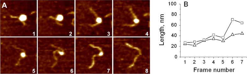
A fundamental challenge associated with chromosomal gene regulation is accessibility of DNA within nucleosomes. Recent studies performed by various techniques, including single-molecule approaches, led to the realization that nucleosomes are dynamic structures rather than static systems, as it was once believed. Direct data is required in order to understand the dynamics of nucleosomes more clearly and answer fundamental questions, including: What is the range of nucleosome dynamics? Does a non-ATP dependent unwrapping process of nucleosomes exist? What are the factors facilitating the large scale opening and unwrapping of nucleosomes? This review summarizes the results of nucleosome dynamics obtained with time-lapse AFM, including a high-speed version (HS-AFM) capable of visualizing molecular dynamics on the millisecond time scale. With HS-AFM, the dynamics of nucleosomes at a sub-second time scale was observed allowing one to visualize various pathways of nucleosome dynamics, such as sliding and unwrapping, including complete dissociation. Overall, these findings reveal new insights into the dynamics of nucleosomes and the novel mechanisms controlling spontaneous chromatin dynamics.

Keywords: Atomic Force Microscopy; chromatin dynamics; nanoimaging; nucleosomes dynamics; single-molecule biophysics.


Figures

Fig. 1
Fig. 1
Schematic explaining the principles of AFM operation. The position of the tip relative to the sample is controlled by a piezoelectric scanner. The vertical displacement of the tip during scanning is detected using the optical lever principle, in which the position of the light spot on the PSPD is measured. The figure was reproduced from Lyubchenko (2011) with permission from Elsevier©
Fig. 2
Fig. 2
AFM image of reconstituted chromatin. The image was taken in air with Tapping mode AFM. AP-mica was used as a substrate for the sample preparation. See Yodh et al. (1999) for specifics
Fig. 3
Fig. 3
Schematics for nucleosome particles with different number of DNA supercoiling turns around the histone core. The figure was reproduced from Shlyakhtenko et al. (2009) with permission from the American Chemical Society©
Fig. 4
Fig. 4
AFM image taken in air of the nucleosome samples. Nucleosomes marked 1, 2, and 3 have about 1.7, 1.4, and 1.0 turns of DNA wrapped around the core particle, respectively. The figure was reproduced from Shlyakhtenko et al. (2009) with permission from the American Chemical Society©
Fig. 5
Fig. 5
Time-lapse AFM of the nucleosome unwrapping process. a Consecutive AFM images of the nucleosome undergoing gradual unwrapping process taken by time-lapse AFM. Each frame size is 200 nm. Each frame takes about 170 s to scan. b Dependence of arm lengths on the frame number. The figure was reproduced from Shlyakhtenko et al. (2009) with permission from the American Chemical Society©
Fig. 6
Fig. 6
Loop formation and unfolding. a A set of images corresponding to 8.7 s (i), 14.7 s (ii) and 17.1 s (iii). In image (ii), the position of the DNA dissociation and unlooping events are indicated with a white arrow. b The length measurements for the looping and the loop unfolding process. The lengths of the left (arm 1) and right (arm 2) arms and the total DNA length are shown with black squares, red diamonds and green triangles, respectively. In (b), the dashed lines correspond to the image acquired times shown in (a). The scan rate is one frame per 301 ms. The figure was reproduced from Miyagi et al. (2011) with permission from the American Chemical Society©
Fig. 7
Fig. 7
Reversible sliding of the nucleosome. a Selected AFM images illustrating a reversible sliding. The vertical dashed line corresponds to the center of nucleosomal particle at 60 s. The numerals (60s, 62s and 65s) correspond to the times the image was captured. b The dependence of the arms’ lengths (arm 1, black squares and arm 2, red diamonds) and the total arms’ lengths (green triangles) on time. The scan rate is 1 frame per second. Scale bar in (a) 50 nm. The figure was reproduced from Lyubchenko (2011), Shlyakhtenko et al. (2009), and Miyagi et al. (2011) with permission from the American Chemical Society©
Fig. 8
Fig. 8
Irreversible sliding of the nucleosome in the presence of CHAPS. ae A set of HS-AFM images. In (a), the black dotted lines indicate the DNA arms of interest. b–e The traced images of the molecule on a black background. f DNA arm length measurements. Black squares and red diamonds correspond to the lengths of the longer and shorter arms in the first image. The green triangles show the time dependence of the total DNA length. The data were acquired with the scan rate of 1.7 frames per second. Dashed white lines in (f) correspond to the times at which images (bd) were acquired, respectively. The figure was reproduced from Miyagi et al. (2011) with permission from the American Chemical Society©

References

    1. Adrian M, Dubochet J, Lepault J, McDowall AW. Cryo-electron microscopy of viruses. Nature. 1984;308(5954):32–36. doi: 10.1038/308032a0. - DOI - PubMed
    1. Ahmad K, Henikoff S. Epigenetic consequences of nucleosome dynamics. Cell. 2002;111(3):281–284. doi: 10.1016/S0092-8674(02)01081-4. - DOI - PubMed
    1. Allen MJ, Dong XF, O’Neill TE, Yau P, Kowalczykowski SC, Gatewood J, Balhorn R, Bradbury EM. Atomic force microscope measurements of nucleosome cores assembled along defined DNA sequences. Biochemistry. 1993;32(33):8390–8396. doi: 10.1021/bi00084a002. - DOI - PubMed
    1. Anderson JD, Thastrom A, Widom J. Spontaneous access of proteins to buried nucleosomal DNA target sites occurs via a mechanism that is distinct from nucleosome translocation. Mol Cell Biol. 2002;22(20):7147–7157. doi: 10.1128/MCB.22.20.7147-7157.2002. - DOI - PMC - PubMed
    1. Bednar J, Horowitz RA, Dubochet J, Woodcock CL. Chromatin conformation and salt-induced compaction: three-dimensional structural information from cryoelectron microscopy. J Cell Biol. 1995;131(6 Pt 1):1365–1376. doi: 10.1083/jcb.131.6.1365. - DOI - PMC - PubMed

LinkOut - more resources