Review
. 2014:2014:197675.
doi: 10.1155/2014/197675. Epub 2014 Apr 15.

Ischemic retinal vasculitis and its management

Affiliations
Review

Ischemic retinal vasculitis and its management

Lazha Talat et al. J Ophthalmol. 2014.

Abstract

Ischemic retinal vasculitis is an inflammation of retinal blood vessels associated with vascular occlusion and subsequent retinal hypoperfusion. It can cause visual loss secondary to macular ischemia, macular edema, and neovascularization leading to vitreous hemorrhage, fibrovascular proliferation, and tractional retinal detachment. Ischemic retinal vasculitis can be idiopathic or secondary to systemic disease such as in Behçet's disease, sarcoidosis, tuberculosis, multiple sclerosis, and systemic lupus erythematosus. Corticosteroids with or without immunosuppressive medication are the mainstay treatment in retinal vasculitis together with laser photocoagulation of retinal ischemic areas. Intravitreal injections of bevacizumab are used to treat neovascularization secondary to systemic lupus erythematosus but should be timed with retinal laser photocoagulation to prevent further progression of retinal ischemia. Antitumor necrosis factor agents have shown promising results in controlling refractory retinal vasculitis excluding multiple sclerosis. Interferon has been useful to control inflammation and induce neovascular regression in retinal vasculitis secondary to Behçet's disease and multiple sclerosis. The long term effect of these management strategies in preventing the progression of retinal ischemia and preserving vision is not well understood and needs to be further studied.


Figures

Figure 1
Figure 1
Histopathological image of a retinal blood vessel involved in Behçet's disease (H & E stain). Note the perivascular infiltration of lymphocytes around the vessel (arrow).
Figure 2
Figure 2
Fundus images of presumed tuberculous occlusive vasculitis. (a) Fundus fluorescein angiography shows peripheral retinal nonperfusion together with small area of hypofluorescence corresponding to chorioretinal lesions along the retinal blood vessels (arrow). (b) A color image showing vascular sheathing together with a fibrovascular tuft originating from the optic disc (arrow), with fluorescein angiography showing (c) leakage at the disc and (d) peripheral capillary dropout and dye leakage from new vessels elsewhere.
Figure 3
Figure 3
Fundus photographs of branch retinal vein occlusion secondary to Behçet's disease. (a, b) Color images of the right eye showing vascular sheathing (arrows), exudates, and intraretinal hemorrhages. (c) Fluorescein angiography demonstrates multiple areas of hypofluorescence corresponding to areas of retinal hemorrhage and (d) upper retinal quadrant hypoperfusion secondary to vasooclusion.
Figure 4
Figure 4
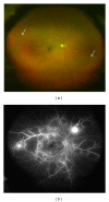
Fundus photographs of SLE associated occlusive retinal vasculitis. (a) Color images demonstrating vascular sheathing (arrows). (b) Fluorescein angiography shows multiple areas of capillary dropout at the retinal midperiphery with leakage from retinal neovascularization (arrows).
Figure 5
Figure 5
Fundus images show vasooclusion in patients with positive anticardiolipin antibodies. (a) Color image showing peripheral branch retinal vein occlusion with an intraretinal haemorrhage and peripheral fibrovascular tuft (Arrow). (b) Fluorescein angiography of the same patient showing vascular fluorescein leakage together with peripheral retinal nonperfusion. (c, d) Fluorescein angiography of another patient demonstrating bilateral retinal ischemia with areas of hypoperfusion covered with laser photocoagulation scars. There are also bilateral fibrovascular tufts leaking fluorescein (arrows).
Figure 6
Figure 6
(a) Color fundus imaging of retinal vasculitis secondary to sarcoidosis showing perivenous exudates, “candle wax drippings.” (b) Fundus fluorescein angiography of an eye with ischemic vasculitis secondary to sarcoidosis shows leakage at macula secondary to macular edema; (c) peripheral retinal hypoperfusion with focal area of fluorescein leakage corresponding to new vessel formation (arrow). (d) Image taken five months following treatment with systemic corticosteroids and focal laser photocoagulation shows regression of the neovascularization.
Figure 7
Figure 7
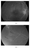
Fundus fluorescein angiography images of a right eye with intermediate uveitis associated with multiple sclerosis; (a) shows fluorescein leakage at the macula secondary to macular edema and (b) peripheral capillary dropout (arrows).
Figure 8
Figure 8
Fundus images of a patient with IRVAN syndrome. (a) Color image showing retinal exudates at the posterior pole involving the macula together with optic disc swelling and hyperemia. (b) Multiple pigmented chorioretinal lesions at the midperiphery (arrow head) together with evidence of vascular sheathing (arrow). (c) Fundus fluorescein angiography showing dye leakage at the optic disc. (d) Wide area of retinal nonperfusion.

References

    1. Rosenbaum JT, Ku J, Ali A, Choi D, Suhler EB. Patients with retinal vasculitis rarely suffer from systemic vasculitis. Seminars in Arthritis and Rheumatism. 2012;41(6):859–865. - PMC - PubMed
    1. Walton RC, Ashmore ED. Retinal vasculitis. Current Opinion in Ophthalmology. 2003;14(6):413–419. - PubMed
    1. Bhaleeya SD, Davis J. Imaging retinal vascular changes in uveitis. International Ophthalmology Clinics. 2012;52(4):83–96. - PubMed
    1. Saurabh K, Das RR, Biswas J, Kumar A. Profile of retinal vasculitis in a tertiary eye care center in Eastern India. Indian Journal of Ophthalmology. 2011;59(4):297–301. - PMC - PubMed
    1. Abu El-Asrar AM, Herbort CP, Tabbara KF. Differential diagnosis of retinal vasculitis. Middle East African Journal of Ophthalmology. 2009;16(4):202–218. - PMC - PubMed