

Transforaminal approach in thoracal disc pathologies: transforaminal microdiscectomy technique
- PMID: 24839557
- PMCID: PMC4009241
- DOI: 10.1155/2014/301945
Transforaminal approach in thoracal disc pathologies: transforaminal microdiscectomy technique
Abstract
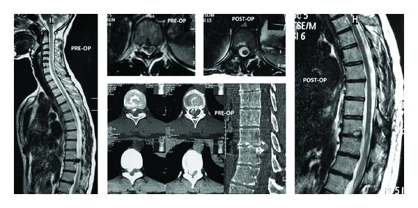
Objective. Many surgical approaches have been defined and implemented in the last few decades for thoracic disc herniations. The endoscopic foraminal approach in foraminal, lateral, and far lateral disc hernias is a contemporary minimal invasive approach. This study was performed to show that the approach is possible using the microscope without an endoscope, and even the intervention on the discs within the spinal canal is possible by having access through the foramen. Methods. Forty-two cases with disc hernias in the medial of the pedicle were included in this study; surgeries were performed with transforaminal approach and microsurgically. Extraforaminal disc hernias were not included in the study. Access was made through the Kambin triangle, foramen was enlarged, and spinal canal was entered. Results. The procedure took 65 minutes in the average, and the mean bleeding amount was about 100cc. They were mobilized within the same day postoperatively. No complications were seen. Follow-up periods range between 5 and 84 months, and the mean follow-up period is 30.2 months. Conclusion. Transforaminal microdiscectomy is a method that can be performed in any clinic with standard spinal surgery equipment. It does not require additional equipment or high costs.
Figures
Similar articles
-
Transforaminal approach in lumbar disc herniations: transforaminal microdiscectomy (TFMD) technique.Turk Neurosurg. 2015;25(1):29-35. doi: 10.5137/1019-5149.JTN.8197-13.1. Turk Neurosurg. 2015. PMID: 25640542
-
Percutaneous Endoscopic Transforaminal Outside-In Outside Technique for Foraminal and Extraforaminal Lumbar Disc Herniations-Operative Technique.World Neurosurg. 2019 Oct;130:244-253. doi: 10.1016/j.wneu.2019.07.005. Epub 2019 Jul 9. World Neurosurg. 2019. PMID: 31299304
-
The Evolution and Advancement of Endoscopic Foraminal Surgery: One Surgeon's Experience Incorporating Adjunctive Techologies.SAS J. 2007 Aug 1;1(3):108-17. doi: 10.1016/SASJ-2006-0014-RR. eCollection 2007. SAS J. 2007. PMID: 25802587 Free PMC article.
-
Operative management of lumbar disc herniation : the evolution of knowledge and surgical techniques in the last century.Acta Neurochir Suppl. 2011;108:17-21. doi: 10.1007/978-3-211-99370-5_4. Acta Neurochir Suppl. 2011. PMID: 21107933 Review.
-
The Evolution of Transforaminal Endoscopic Spine Surgery.World Neurosurg. 2021 Jan;145:643-656. doi: 10.1016/j.wneu.2020.08.096. Epub 2020 Aug 19. World Neurosurg. 2021. PMID: 32822954 Review.
Cited by
-
Minimally invasive transpedicular approach for the treatment of central calcified thoracic disc disease: a technical note.Eur Spine J. 2018 Jul;27(7):1575-1585. doi: 10.1007/s00586-017-5406-y. Epub 2017 Dec 15. Eur Spine J. 2018. PMID: 29247397
-
Phase I 270° single-incision percutaneous spinal endoscopy for decompression treatment of thoracic spinal stenosis.Sci Rep. 2022 Jun 8;12(1):9448. doi: 10.1038/s41598-022-13666-4. Sci Rep. 2022. PMID: 35676323 Free PMC article.
-
Percutaneous Discectomy Followed by CESI Might Improve Neurological Disorder of Drop Foot Patients Due to Chronic LDH.Brain Sci. 2020 Aug 11;10(8):539. doi: 10.3390/brainsci10080539. Brain Sci. 2020. PMID: 32796497 Free PMC article.
References
-
- Bransford R, Zhang F, Bellabarb C, Konodi M, Chapman JR. Early experience treating thoracic disc herniations using a modified transfacet pedicle-sparing decompression and fusion: clinical article. Journal of Neurosurgery: Spine. 2010;12(2):221–231. - PubMed
-
- Stillerman CB, Chen TC, Day JD, Couldwell WT, Weiss MH. The transfacet pedicle-sparing approach for thoracic disc removal: cadaveric morphometric analysis and preliminary clinical experience. Journal of Neurosurgery. 1995;83(6):971–976. - PubMed
-
- Ross JS, Perez-Reyes N, Masaryk TJ, Bohlman H, Modic MT. Thoracic disk herniation: MR imaging. Radiology. 1987;165(2):511–515. - PubMed
-
- Arseni C, Nash F. Thoracic intervertebral disc protrusion: a clinical study. Journal of Neurosurgery. 1960;17:418–430. - PubMed
-
- Uribe JS, Smith WD, Pimenta L, et al. Minimally invasive lateral approach for symptomatic thoracic disc herniation: initial multicenter clinical experience—clinical article. Journal of Neurosurgery: Spine. 2012;16(3):264–279. - PubMed
LinkOut - more resources
Full Text Sources
Other Literature Sources
