Case Reports
. 2014 Jul;128(1):111-22.
doi: 10.1007/s00401-014-1285-z. Epub 2014 May 20.

Pituitary blastoma: a pathognomonic feature of germ-line DICER1 mutations

Affiliations
Case Reports

Pituitary blastoma: a pathognomonic feature of germ-line DICER1 mutations

Leanne de Kock et al. Acta Neuropathol. 2014 Jul.

Abstract

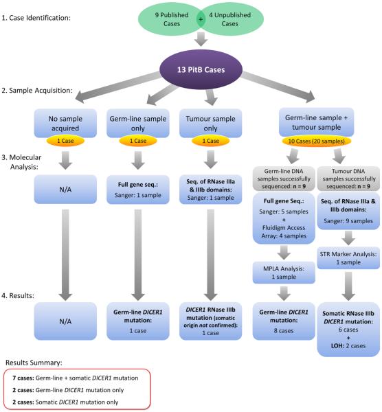
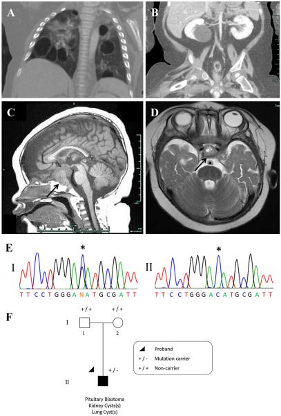
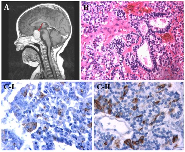
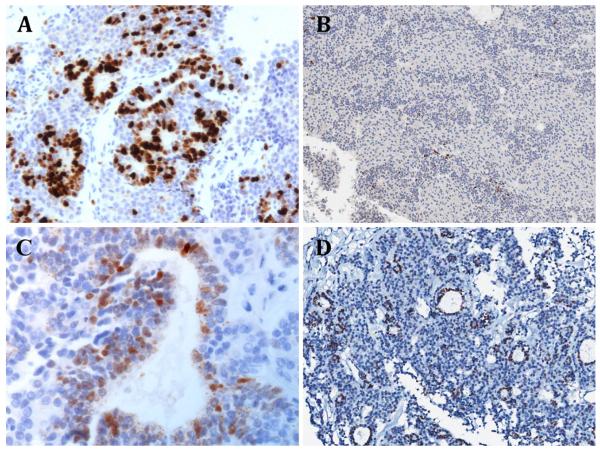
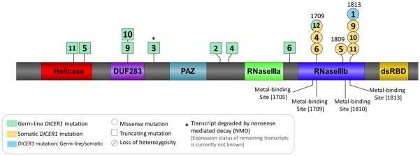
Individuals harboring germ-line DICER1 mutations are predisposed to a rare cancer syndrome, the DICER1 Syndrome or pleuropulmonary blastoma-familial tumor and dysplasia syndrome [online Mendelian inheritance in man (OMIM) #601200]. In addition, specific somatic mutations in the DICER1 RNase III catalytic domain have been identified in several DICER1-associated tumor types. Pituitary blastoma (PitB) was identified as a distinct entity in 2008, and is a very rare, potentially lethal early childhood tumor of the pituitary gland. Since the discovery by our team of an inherited mutation in DICER1 in a child with PitB in 2011, we have identified 12 additional PitB cases. We aimed to determine the contribution of germ-line and somatic DICER1 mutations to PitB. We hypothesized that PitB is a pathognomonic feature of a germ-line DICER1 mutation and that each PitB will harbor a second somatic mutation in DICER1. Lymphocyte or saliva DNA samples ascertained from ten infants with PitB were screened and nine were found to harbor a heterozygous germ-line DICER1 mutation. We identified additional DICER1 mutations in nine of ten tested PitB tumor samples, eight of which were confirmed to be somatic in origin. Seven of these mutations occurred within the RNase IIIb catalytic domain, a domain essential to the generation of 5p miRNAs from the 5' arm of miRNA-precursors. Germ-line DICER1 mutations are a major contributor to PitB. Second somatic DICER1 "hits" occurring within the RNase IIIb domain also appear to be critical in PitB pathogenesis.


Figures

Fig. 1
Fig. 1
Flowchart summarizing case identification, sample acquisition, molecular analysis and results of the study
Fig. 2
Fig. 2
Case 12: a chest CT following IV contrast: multiple bilateral thin-walled air filled cysts evident within all lobes of the lungs; likely PPB Type I or Ir (not biopsied). b Coronal CT image of abdomen and pelvis. Liquid-filled cyst in lower pole of right kidney is typical of CN (not biopsied). Other CT images (not shown) also revealed a smaller cyst (likely CN, not biopsied) in the left kidney. c T1 midline sagittal MR image. d T2-weighted axial MR image just superior to the pituitary. In c and d, the pituitary tumor is indicated with arrows. e Panel I a mono-allelic germ-line DICER1 mutation, c.5125G>C [p.(Asp1709His)]. Panel II clear loss of heterozygosity at the position of the germ-line DICER1 mutation within the tumor (wild-type allele lost). f The proband (individual II-1) was diagnosed with PitB at the age of 8 months and was found to carry the de novo c.5125G>C germ-line DICER1 mutation
Fig. 3
Fig. 3
a case 13, T1-weighted post-contrast midline sagittal MR image showing pituitary region mass (red arrow). b case 4, hematoxylin and eosin (H&E) staining ×250: three enlarged follicles lined by stem cells. c Immunohistochemical staining I case 10, Growth hormone (GH) immunostaining ×400: enlarged GH/alpha subunit cells immunopositive for GH. II case 10, ACTH immunostaining ×400: small vessel surrounded by stem cells. Some cells display ACTH immunoreactivity
Fig. 4
Fig. 4
Immunohistochemical staining a case 12, Ki-67 labelling ×200: there is marked labelling for Ki-67, indicating a high proliferative fraction that is limited to the rosette-like epithelial structures. b Case 2, Ki-67 labelling ×100: Ki-67 labelling index estimated at 1.63 %, indicating low proliferative activity. c Case 12, p53 immunostaining ×400. d Case 2, p53 immunostaining ×100. For c and d, p53 expression is present in cells forming the rosette structures, but is scant elsewhere
Fig. 5
Fig. 5
Graphic representation of the DICER1 protein structure (NP_001258211.1) indicating the approximate positions of the germ-line (green diagonal stripes) and somatic (orange horizontal stripes) DICER1 mutations observed in the 13 cases being reported. Mutation shaded with blue vertical stripes represent mutations that were identified within tumour gDNA, but are not confirmed to be somatic in origin. Case number indicated at the position of each mutation: case 1: somatic DICER1 amino acid change: p.(Glu1813Lys). Case 2: germ-line DICER1 amino acid change: p.(Asn1093*). Case 3: germ-line DICER1 amino acid change: p.(Tyr793*), (NMD of mutant). Case 4: germ-line DICER1 amino acid change: p.(Ser1179Thrfs*12); Somatic DICER1 amino acid change: p.(Asp1709Thr). Case 5: germ-line DICER1 amino acid change: p.(Arg509*); somatic DICER1 amino acid change: p.(Gly1809Trp). Case 6: germ-line DICER1 amino acid change: p.(Asp1437Metfs*16); somatic DICER1 amino acid change: p.(Asp1709Asn). Case 9: germ-line DICER1 amino acid change: p.(Arg676*); somatic DICER1 amino acid change: p.(Glu1813Asp). Case 10: germ-line DICER1 amino acid change: p.(Arg676*); somatic DICER1 amino acid change: p.(Glu1813Val). Case 11: germ-line DICER1 amino acid change: p.(Lys429Alafs*47); somatic DICER1 amino acid change: p.(Glu1813Lys). Case 12: germ-line DICER1 amino acid change: p.(Asp1709His); somatic DICER1 change: loss of heterozygosity

References

    1. Anglesio MS, Wang Y, Yang W, Senz J, Wan A, Heravi-Moussavi A, Salamanca C, Maines-Bandiera S, Huntsman DG, Morin GB. Cancer-associated somatic DICER1 hotspot mutations cause defective miRNA processing and reverse-strand expression bias to predominantly mature 3p strands through loss of 5p strand cleavage. J Pathol. 2013;229(3):400–409. doi:10.1002/path.4135. - PubMed
    1. Bahubeshi A, Bal N, Rio Frio T, Hamel N, Pouchet C, Yilmaz A, Bouron-Dal Soglio D, Williams GM, Tischkowitz M, Priest JR, Foulkes WD. Germline DICER1 mutations and familial cystic nephroma. J Med Genet. 2010;47(12):863–866. doi:10.1136/jmg.2010.081216. - PubMed
    1. Baker M. Cancer stem cells tracked. Nature. 2012;488(7409):13–14. doi:10.1038/488013a. - PubMed
    1. Chen J, Li Y, Yu TS, McKay RM, Burns DK, Kernie SG, Parada LF. A restricted cell population propagates glioblastoma growth after chemotherapy. Nature. 2012;488(7412):522–526. doi:10.1038/nature11287. - PMC - PubMed
    1. Foulkes WD, Bahubeshi A, Hamel N, Pasini B, Asioli S, Baynam G, Choong CS, Charles A, Frieder RP, Dishop MK, Graf N, Ekim M, Bouron-Dal Soglio D, Arseneau J, Young RH, Sabbaghian N, Srivastava A, Tischkowitz MD, Priest JR. Extending the phenotypes associated with DICER1 mutations. Hum Mutat. 2011;32(12):1381–1384. doi:10.1002/humu.21600. - PubMed

Publication types

MeSH terms