

Computational fluid dynamics endpoints for assessment of adenotonsillectomy outcome in obese children with obstructive sleep apnea syndrome
- PMID: 24840295
- PMCID: PMC4057952
- DOI: 10.1016/j.jbiomech.2014.03.023
Computational fluid dynamics endpoints for assessment of adenotonsillectomy outcome in obese children with obstructive sleep apnea syndrome
Abstract
Background: Improvements in obstructive sleep apnea syndrome (OSAS) severity may be associated with improved pharyngeal fluid mechanics following adenotonsillectomy (AT). The study objective is to use image-based computational fluid dynamics (CFD) to model changes in pharyngeal pressures after AT, in obese children with OSAS and adenotonsillar hypertrophy.
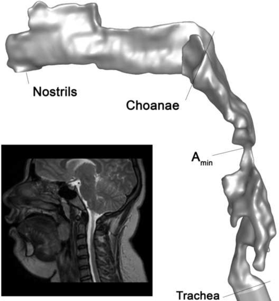
Methods: Three-dimensional models of the upper airway from nares to trachea, before and after AT, were derived from magnetic resonance images obtained during wakefulness, in a cohort of 10 obese children with OSAS. Velocity, pressure, and turbulence fields during peak tidal inspiratory flow were computed using commercial software. CFD endpoints were correlated with polysomnography endpoints before and after AT using Spearman׳s rank correlation (rs).
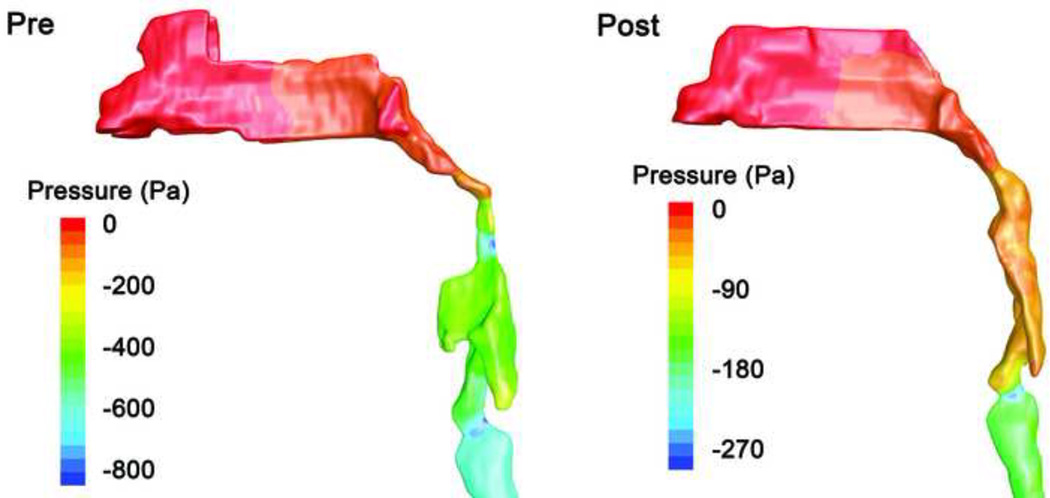
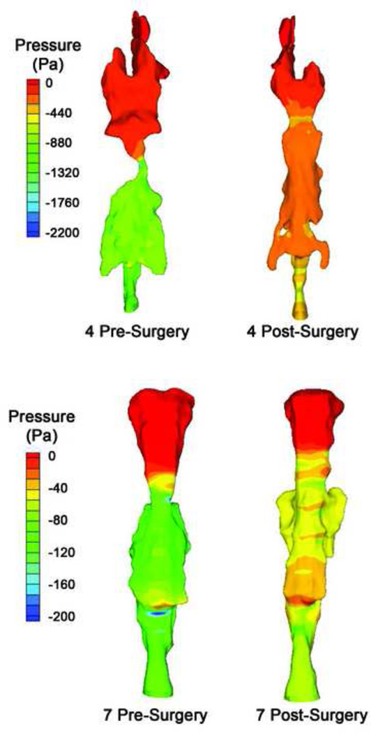
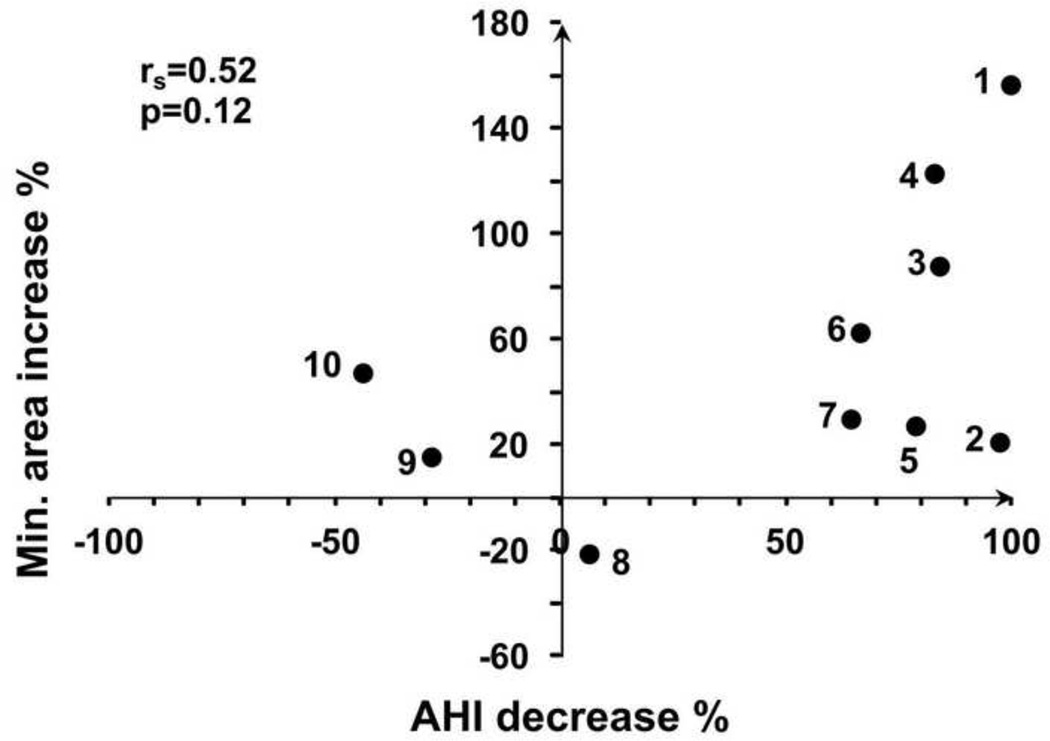
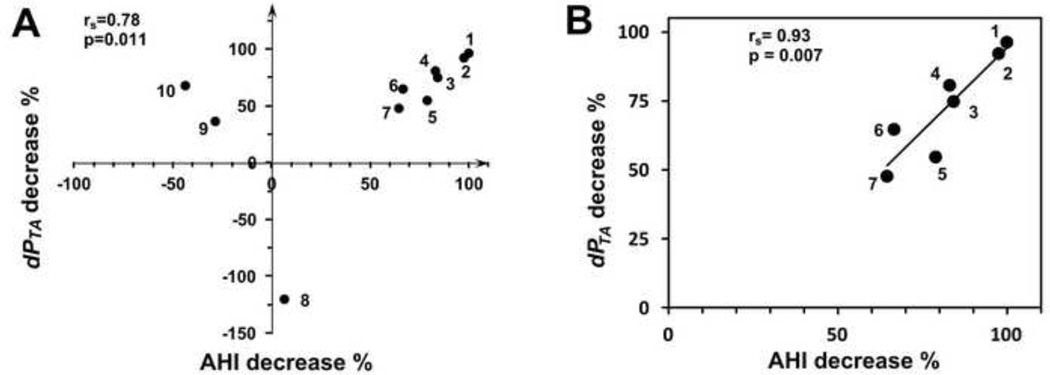
Results: Apnea hypopnea index (AHI) decreases after AT was strongly correlated with reduction in maximum pressure drop (dPTAmax) in the region where tonsils and adenoid constrict the pharynx (rs=0.78, P=0.011), and with decrease of the ratio of dPTAmax to flow rate (rs=0.82, P=0.006). Correlations of AHI decrease to anatomy, negative pressure in the overlap region (including nasal flow resistance), or pressure drop through the entire pharynx, were not significant. In a subgroup of subjects with more than 10% improvement in AHI, correlations between flow variables and AHI decrease were stronger than in all subjects.
Conclusions: The correlation between change in dPTAmax and improved AHI suggests that dPTAmax may be a useful index for internal airway loading due to anatomical narrowing, and may be better correlated with AHI than direct airway anatomic measurements.
Keywords: Airway resistance; Computer simulation; Humans; Magnetic resonance imaging; Pediatrics.
Copyright © 2014 Elsevier Ltd. All rights reserved.
Figures
References
-
- American Academy of Pediatrics, So. P.P., Subcommittee on Obstructive Sleep Apnea Syndrome. Clinical practice guideline: diagnosis and management of childhood obstructive sleep apnea syndrome. Pediatrics. 2002;109:704–712. - PubMed
-
- Arens R, McDonough JM, Corbin AM, Hernandez EM, Maislin G, Schwab RJ, Pack AI. Linear dimensions of the upper airway structure during development: assessment by magnetic resonance imaging. Am J Respir Crit Care Med. 2002;165:117–122. - PubMed
-
- Arens R, McDonough JM, Corbin AM, Rubin NK, Carroll ME, Pack AI, Liu J, Udupa JK. Upper airway size analysis by magnetic resonance imaging of children with obstructive sleep apnea syndrome. Am J Respir Crit Care Med. 2003;167:65–70. - PubMed
-
- Arens R, McDonough JM, Costarino AT, Mahboubi S, Tayag-Kier CE, Maislin G, Schwab RJ, Pack AI. Magnetic resonance imaging of the upper airway structure of children with obstructive sleep apnea syndrome. Am J Respir Crit Care Med. 2001;164:698–703. - PubMed
-
- Block AJ, Faulkner JA, Hughes RL, Remmers JE, Thach B. Clinical conference in pulmonary disease. Factors influencing upper airway closure. Chest. 1984;86:114–122. - PubMed
Publication types
MeSH terms
Grants and funding
LinkOut - more resources
Full Text Sources
Other Literature Sources
Medical
Miscellaneous
