. 2014 Jul 15;74(14):3923-34.
doi: 10.1158/0008-5472.CAN-13-1866. Epub 2014 May 19.

Inactivation of p53 is insufficient to allow B cells and B-cell lymphomas to survive without Dicer

Affiliations

Inactivation of p53 is insufficient to allow B cells and B-cell lymphomas to survive without Dicer

Clare M Adams et al. Cancer Res. .

Abstract

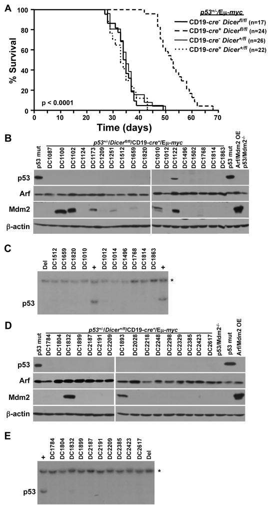
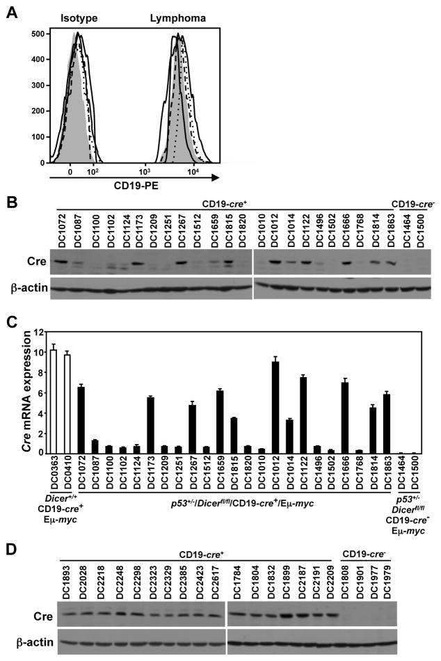
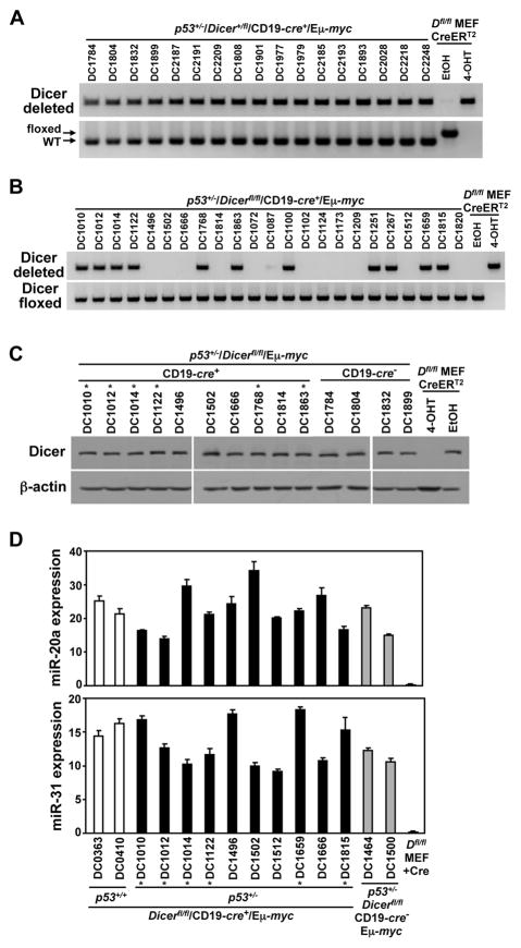
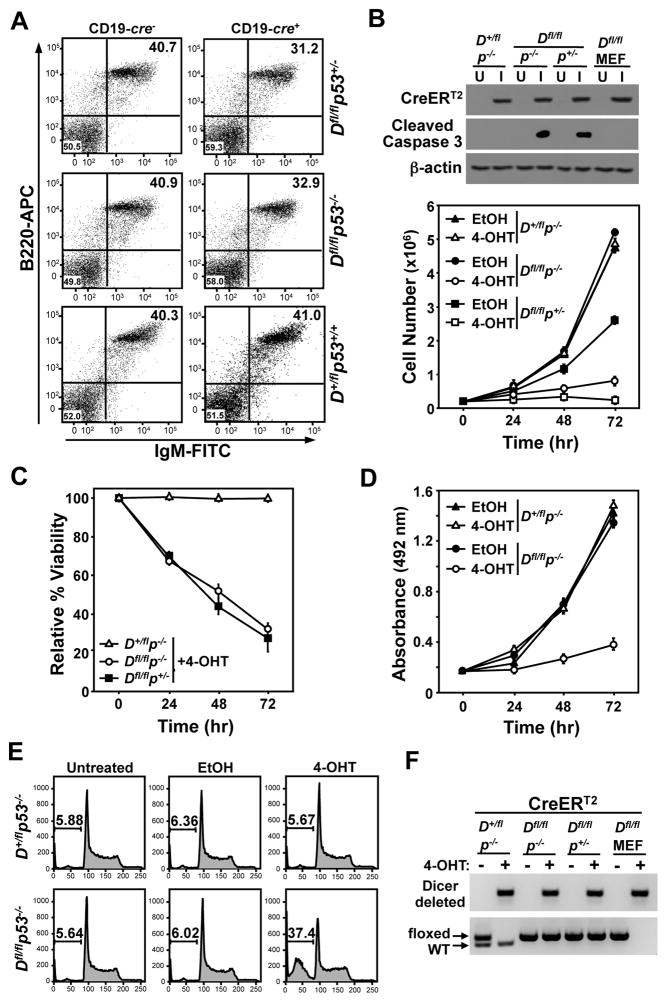
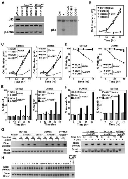
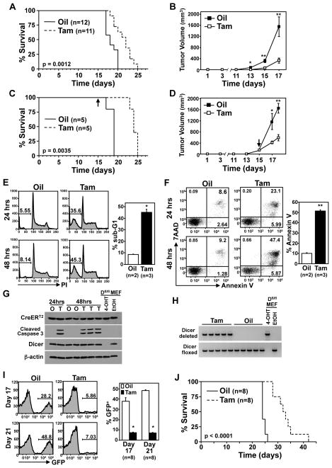
Inactivation of p53, the master regulator of cellular stress and damage signals, often allows cells that should die or senesce to live. Loss of Dicer, an RNase III-like enzyme critical in microRNA biogenesis, causes embryonic lethality and activation of the p53 pathway. Several nonhematopoietic cell types that contain inactivated p53 have been shown to survive Dicer deletion, suggesting that p53 loss may protect cells from the negative consequences of Dicer deletion. However, here, we report that loss of p53 did not provide a survival advantage to B cells, as they underwent rapid apoptosis upon Dicer deletion. Moreover, a deficiency in p53 neither rescued the Dicer deletion-induced delay in Myc-driven B-cell lymphomagenesis, nor allowed a single B-cell lymphoma to develop with biallelic deletion of Dicer. A p53 deficiency did, however, restore the pre-B/B-cell phenotype and CD19 surface expression of the lymphomas that emerged in conditional Dicer knockout Eμ-myc transgenic mice. Moreover, p53 loss in transformed B cells did not confer protection from apoptosis, as Dicer deletion in established p53-null B-cell lymphomas induced apoptosis, and all of the 1,260 B-cell lymphoma clones analyzed that survived Cre-mediated Dicer deletion retained at least one allele of Dicer. Moreover, Dicer deletion in lymphomas in vivo reduced tumor burden and prolonged survival. Therefore, inactivation of p53 is insufficient to allow untransformed B cells and B-cell lymphomas to survive without Dicer, presenting a potential therapeutic opportunity for the treatment of B-cell lymphomas.


Conflict of interest statement

The authors declare no conflict of interest

Figures

Figure 1
Figure 1. Delayed lymphomagenesis in p53+/−/CD19-cre+/Dicerfl/fl/Eμ-myc mice
(A) Kaplan-Meier survival curves of the indicated genotypes of mice (p<0.0001, log-rank test comparing each genotype to CD19-cre+/Dicerfl/fl/p53+/−/Eμ-myc). The number (n) of mice is indicated. (B, D) Western blots of lymphomas for the proteins and genotype indicated. Controls include lymphomas containing mutant (mut) p53 or overexpressing (OE) Arf and Mdm2 and p53−/−/Mdm2−/− MEFs. A subset of lymphomas analyzed shown. (C, E) Representative Southern blots for p53 of lymphomas in B and D. Lymphomas that contain (+) or have deleted (Del) p53 were controls. Asterisk (*) denotes the DNA loading control, the p53 pseudogene.
Figure 2
Figure 2. A deficiency in p53 rescues CD19 and Cre expression during B-cell lymphomagenesis
(A) Histograms of CD19 surface expression and corresponding isotype controls of lymphomas from four representative p53+/−/CD19-cre+/Dicerfl/fl/Eμ-myc mice compared to a control p53+/−/CD19-cre/Dicerfl/fl/Eμ-myc lymphoma (shaded peaks). (B, D) Western blots for Cre and β-actin from Dicerfl/fl (B) and Dicer+/fl (D) p53+/−/CD19-cre+/Eμ-myc lymphomas. Lysates of lymphomas from a Dicer+/+/CD19-cre+/Eμ-myc mouse (B) and Dicerfl/fl (B) or Dicer+/fl (D) CD19-cre/p53+/−/Eμ-myc mice were controls. (C) qRT-PCR for Cre expression relative to β-actin in lymphomas from p53+/−/CD19-cre+/Dicerfl/fl/Eμ-myc mice. RNA from Dicer+/+/CD19-cre+/Eμ-myc and CD19-cre/p53+/−/Dicerfl/fl/Eμ-myc lymphomas were positive and negative controls, respectively.
Figure 3
Figure 3. Biallelic Dicer deletion is selected against during lymphoma development
(A, B) PCR analysis for conditional deleted and floxed (not deleted) Dicer alleles from lymphomas of the indicated genotype. DNA from Dicerfl/fl (Dfl/fl) MEFs expressing an inducible CreERT2 treated with 4-OHT or vehicle control (EtOH) were controls. Arrows indicate unrearranged (floxed) and wild-type (WT) Dicer alleles. (C) Representative Western blots for Dicer and β-actin from CD19-cre+ and CD19-cre p53+/−/Dicerfl/fl/Eμ-myc lymphomas. Lysates from Dicerfl/fl (Dfl/fl) MEFs were controls. (D) qRT-PCR for miR-20a and miR-31 relative to internal RNU6b small RNA in lymphomas from p53+/−/CD19-cre+/Dicerfl/fl/Eμ-myc mice. p53+/+/CD19-cre+/Dicerfl/fl/Eμ-myc and p53+/−/Dicerfl/fl/CD19-cre/Eμ-myc lymphomas and Cre-expressing Dicerfl/fl (Dfl/fl) MEFs served as controls. Asterisks (*) denote lymphomas that deleted one Dicer allele (C and D).
Figure 4
Figure 4. Loss of p53 is insufficient for B-cell survival when Dicer is deleted
(A) Representative dot plots of littermate-matched splenic B-cells from CD19-cre+ or CD19-cre Dicer+/fl and Dicerfl/fl mice that were p53+/+, p53+/−, or p53−/−. Total lymphocytes were gated and B220-APC versus IgM-FITC was assessed. (B–F) Primary pre-B cells from p53−/−/Dicerfl/fl, p53+/−/Dicerfl/fl, and p53−/−/Dicer+/fl littermates were infected (I) with a retrovirus encoding CreERT2 or left uninfected (U). 4-OHT (+) or vehicle control (EtOH, −) was added to pre-B cell cultures at time 0 and cell number (B), viability (C), proliferation (MTS assay; D), apoptosis (cleaved Caspase 3 protein, B; sub-G1 DNA, E), and Dicer gene rearrangement (F) were evaluated. Western blots shown in B. Arrows indicate unrearranged (floxed) and wild-type (WT) Dicer alleles in F. CreERT2 expressing Dicerfl/fl (Dfl/fl) MEFs treated with 4-OHT (+) or ethanol (−) were controls in B and F.
Figure 5
Figure 5. A deficiency in p53 does not allow B-cell lymphomas to survive without Dicer
(A) p53+/−/Dicerfl/fl/Eμ-myc (DC1020 and DC1185), p53+/−/Dicer+/fl/Eμ-myc (DC2385 and DC2423) lymphoma cell lines and the Dicerfl/fl/Eμ-myc lymphoma cell line (DC561) from our previous study (19) were subjected to Western blot (left) for the proteins indicated and Southern blot (right and Fig. 1E) for p53. A lymphoma containing mutant p53 was a control for the Western blot. Lymphomas that contain (+) or have deleted (Del) p53 were controls for the Southern blot. Asterisk (*) denotes the DNA loading control, the p53 pseudogene. (B–F) DC2385, DC2423, DC1020, and/or DC1185 lymphoma cells were infected with a CreERT2-encoding retrovirus or empty retrovirus (Vector). 4-OHT or vehicle control (EtOH) was added to the cultures at time 0 and cell number (B, C), viability (D), and apoptosis (sub-G1 DNA, E; Annexin V, F) were measured. (G) Dicer gene rearrangement was evaluated at the indicated intervals by PCR. (H) Representative PCR product analysis of Dicer gene rearrangement of GFP-positive single cell-sorted lymphoma clones that survived CreERT2 activation of the 328 analyzed. Conditional deleted and floxed (not deleted) Dicer alleles shown (G and H). CreERT2 expressing Dicerfl/fl (Dfl/fl) MEFs treated with 4-OHT or ethanol (EtOH) were controls (G and H).
Figure 6
Figure 6. Dicer inactivation impedes tumor growth, in vivo
(A, C) Kaplan-Meier survival curves of nude mice injected (subcutaneously) with CreERT2 expressing p53 deleted Dicerfl/fl/Eμ-myc lymphoma cells (DC1020) and administered tamoxifen or vehicle (corn oil) control starting the day of injection (A; p=0.0012, log-rank test) or once lymphomas were 90–150mm3 (C; p=0.0035, log-rank test). Arrow indicates the day tamoxifen administration began for C. The number (n) of mice is indicated. (B, D) Tumor volumes for mice in A and C, respectively, were measured at the indicated intervals (for B: *p=0.0051, **p<0.003; for D: *p=0.0288, **p=0.0005). In D, the arrow indicates the day tamoxifen administration began. (E–G) Apoptosis was measured at intervals following tamoxifen or vehicle control administration in matched tumor pairs by propidium iodide staining of fragmented (sub-G1) DNA (E), Annexin V/7AAD staining (F), and cleaved Caspase 3 protein detection (G). Representative data (left) and mean values at 48 hours (right) are shown for E and F; *p=0.0008, **p<0.0001, t-tests. Western blots of whole cell lysates for the proteins indicated (G). (H) PCR product analysis of Dicer gene rearrangement of the mice from C. Controls for G and H include protein lysates or DNA from Dicerfl/fl MEFs treated with 4-OHT or ethanol. (I, J) Nude mice were injected intravenously with CreERT2 expressing p53 deleted Dicerfl/fl/Eμ-myc lymphoma cells (DC1020) and administered tamoxifen (Tam) or corn oil (Oil) vehicle control starting the same day. Blood was assessed for GFP-positivity by flow cytometry at intervals post lymphoma injection. Representative data (left) and mean values for the indicated number of mice are shown (I; *p<0.0001, t-test). Kaplan-Meier survival curves (J; p<0.0001, log-rank test).

References

    1. Almeida MI, Reis RM, Calin GA. MicroRNA history: discovery, recent applications, and next frontiers. Mutat Res. 2011;717:1–8. - PubMed
    1. Mendell JT, Olson EN. MicroRNAs in stress signaling and human disease. Cell. 2012;148:1172–87. - PMC - PubMed
    1. Iorio MV, Croce CM. microRNA involvement in human cancer. Carcinogenesis. 2012;33:1126–33. - PMC - PubMed
    1. Bui TV, Mendell JT. Myc: Maestro of MicroRNAs. Genes Cancer. 2010;1:568–75. - PMC - PubMed
    1. Finnegan EF, Pasquinelli AE. MicroRNA biogenesis: regulating the regulators. Crit Rev Biochem Mol Biol. 2013;48:51–68. - PMC - PubMed

Publication types

MeSH terms