. 2014 Jul 11;289(28):19571-84.
doi: 10.1074/jbc.M114.550624. Epub 2014 May 19.

Mitochondrial NLRP3 protein induces reactive oxygen species to promote Smad protein signaling and fibrosis independent from the inflammasome

Affiliations

Mitochondrial NLRP3 protein induces reactive oxygen species to promote Smad protein signaling and fibrosis independent from the inflammasome

Nathan A Bracey et al. J Biol Chem. .

Abstract

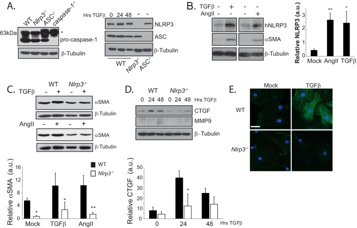
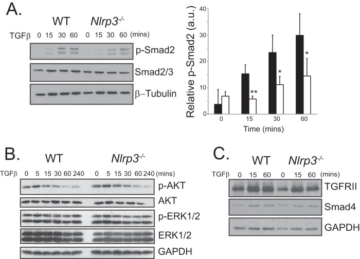
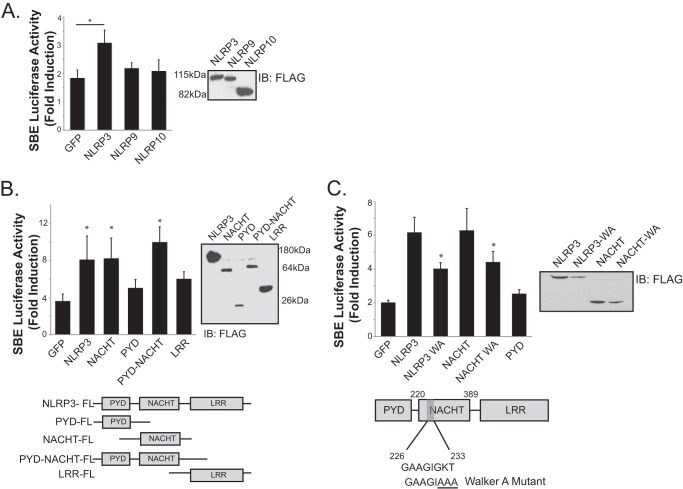
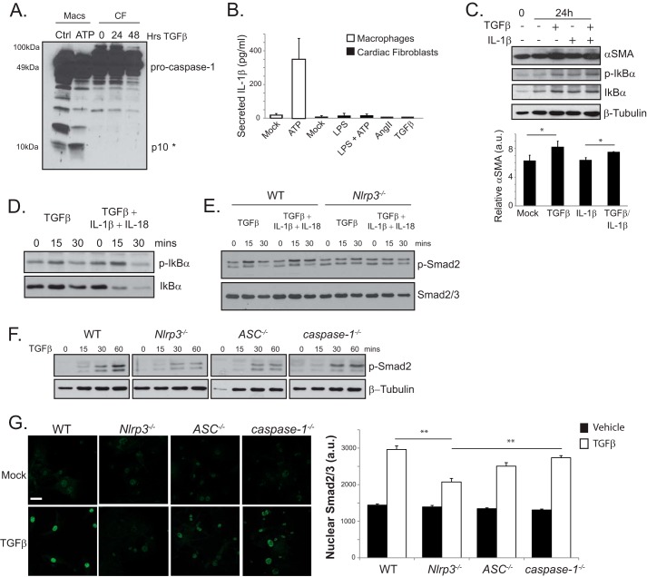
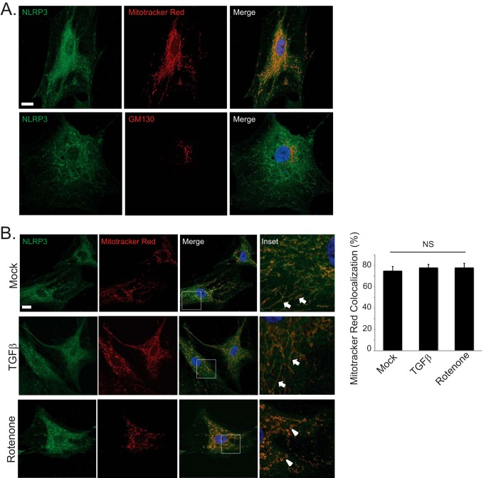
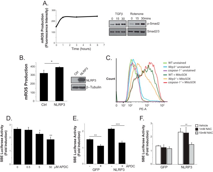
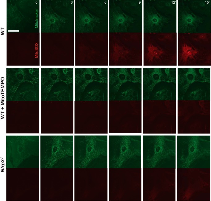
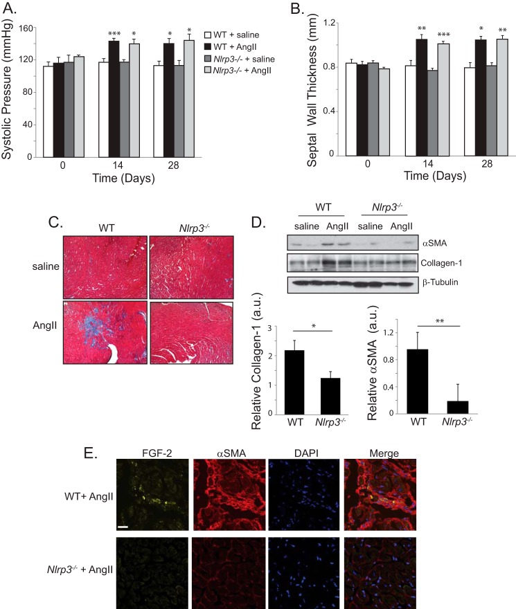
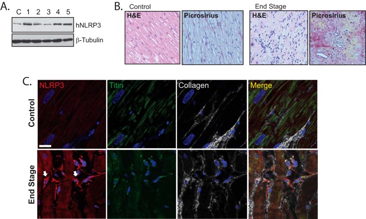
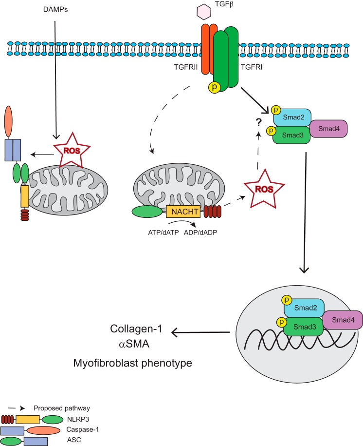
Nucleotide-binding domain and leucine-rich repeat containing PYD-3 (NLRP3) is a pattern recognition receptor that is implicated in the pathogenesis of inflammation and chronic diseases. Although much is known regarding the NLRP3 inflammasome that regulates proinflammatory cytokine production in innate immune cells, the role of NLRP3 in non-professional immune cells is unclear. Here we report that NLRP3 is expressed in cardiac fibroblasts and increased during TGFβ stimulation. NLRP3-deficient cardiac fibroblasts displayed impaired differentiation and R-Smad activation in response to TGFβ. Only the central nucleotide binding domain of NLRP3 was required to augment R-Smad signaling because the N-terminal Pyrin or C-terminal leucine-rich repeat domains were dispensable. Interestingly, NLRP3 regulation of myofibroblast differentiation proceeded independently from the inflammasome, IL-1β/IL-18, or caspase 1. Instead, mitochondrially localized NLRP3 potentiated reactive oxygen species to augment R-Smad activation. In vivo, NLRP3-deficient mice were protected against angiotensin II-induced cardiac fibrosis with preserved cardiac architecture and reduced collagen 1. Together, these results support a distinct role for NLRP3 in non-professional immune cells independent from the inflammasome to regulate differential aspects of wound healing and chronic disease.

Keywords: Fibrosis; Heart Failure; Inflammation; Myofibroblast; Transforming Growth Factor β (TGFβ).


Figures

FIGURE 1.
FIGURE 1.
NLRP3 regulates human cardiac myofibroblast differentiation. A, immunoblotting for NLRP3, ASC, and pro-caspase 1 in murine CFs. The asterisk denotes a nonspecific band present at 63kDa. B, immunoblotting and semiquantitative analysis for NLRP3 and αSMA in human CFs (hNLRP3) stimulated for 24 h with TGFβ (10 ng/ml) or AngII (1 μm). a.u., arbitrary units. *, p < 0.05; **, p < 0.01. C and D, immunoblotting for CTGF, MMP9, and αSMA in WT and Nlrp3−/− CFs following stimulation with TGFβ or AngII (n = 4). *, p < 0.05; **, p < 0.01. E, confocal fluorescent immunocytochemistry of αSMA in WT and Nlrp3−/− CFs following 24-h stimulation with TGFβ. Scale bar = 10 μm.
FIGURE 2.
FIGURE 2.
NLRP3 regulates Smad signaling. A, immunoblotting and semiquantitative analysis for phospho-Smad2 (p-Smad2) in TGFβ-stimulated WT and Nlrp3−/− CFs for the indicated time points (n = 4). *, p < 0.05; **, p < 0.01. B and C, immunoblotting for phospho-ERK1/2 (p-ERK1/2), phospho-AKT (p-AKT), TGFRII, and Smad4 in WT and Nlrp3−/− CFs stimulated with TGFβ at the indicated time points.
FIGURE 3.
FIGURE 3.
The NLRP3 NACHT domain is required for promotion of Smad signaling. A, luciferase assay and expression of FLAG-tagged NLRP3, NLRP9, and NLRP10 constructs with the SBE4-luciferase reporter in 293T cells transfected with control GFP or NLRs. IB, immunoblot. B, luciferase assay in 293T cells transfected with control GFP, NLRP3, NACHT, PYD, PYD-NACHT, or LRR constructs. Data are expressed as fold induction of luciferase activity following TGFβ stimulation (10 ng/ml) compared with mock-treated cells (n = 3). *, p < 0.05 versus GFP. C, structure and expression of the NLRP3 Walker A (WA) mutation in full-length NLRP3 and NACHT constructs. SBE4-luciferase assay with control GFP, NLRP3, NLRP3 Walker A mutant, NACHT, NACHT Walker A mutant, or PYD constructs (n = 3). *, p < 0.05 versus NLRP3.
FIGURE 4.
FIGURE 4.
Inflammasome independence of NLRP3 in TGFβ-induced Smad signaling. A, immunoblotting for caspase 1 processing in WT CFs stimulated for 24 and 48 h with TGFβ. Primary murine peritoneal macrophages (Macs) primed with LPS and stimulated with 5 mm ATP for 1 h served as a positive control (Ctrl). The asterisk denotes the cleaved caspase 1 p10 band. B, IL-1β in supernatants from WT CFs primed with LPS (10 ng/ml) followed by ATP (5 mm) or stimulated with AngII or TGFβ. C, immunoblotting and semiquantitative analysis for αSMA and phospho-IκBα (p-IκBα) in fibroblasts stimulated with TGFβ, IL-1β (1 ng/ml), or both (n = 3). a.u., arbitrary units. *, p < 0.05. D and E, immunoblotting for phospho-IκBα in Nlrp3−/− cardiac fibroblasts and phospho-Smad2 (p-Smad2) in WT and Nlrp3−/− CFs stimulated with TGFβ, IL-1β/IL-18 (1 ng/ml and 10 ng/ml, respectively), or all three. F, immunoblot for phospho-Smad2 in WT, Nlrp3−/−, ASC−/−, and caspase 1−/− CFs stimulated with TGFβ. G, confocal immunofluorescence and quantification of nuclear Smad2/3 in WT, Nlrp3−/−, ASC−/−, and caspase 1−/− CFs stimulated with TGFβ for 15 min. **, p < 0.01; n > 30 cells in at least four fields of view. Error bars indicate mean ± S.E. Scale bar = 40 μm.
FIGURE 5.
FIGURE 5.
Mitochondrial localization of endogenous NLRP3 in human cardiac fibroblasts. A, confocal fluorescent immunocytochemistry for endogenous NLRP3 and MitoTracker Red (top row) or the cis-Golgi marker GM130 (bottom row) in unstimulated human CFs. Scale bar = 20 μm. B, confocal fluorescent immunocytochemistry for endogenous NLRP3 and MitoTracker Red in human CFs stimulated with TGFβ for 24 h or rotenone (10 μm) for 6 h. Solid arrows are directed at mitochondria with a linear morphology, and arrowheads are directed at mitochondria with a fragmented morphology. Scale bar = 20 μm. NS, not significant.
FIGURE 6.
FIGURE 6.
Role of mitochondrial ROS in NLRP3-mediated Smad signaling. A, left panel, mROS measurements on MitoSox-loaded (5 μm) 293T cells transfected with the TGF type II receptor, stimulated with TGFβ for the indicated times, and subjected to flow cytometry. Data are expressed as mean fluorescence intensity units. Right panel, immunoblot for phosphorylated Smad2 (p-Smad2) in human CFs treated with either TGFβ or rotenone (10 μm). B, flow cytometry quantification of MitoSox-loaded (5 μm) 293T cells transfected with either a control plasmid (Ctrl) or NLRP3. *, p < 0.05; n = 3. C, flow cytometry on control and MitoSox-loaded WT, Nlrp3−/−, and caspase 1−/− CFs. D and E, luciferase assay with SBE4-luciferase reporter in 293T cells transfected with NLRP3 and stimulated with TGFβ in the presence of increasing concentrations of aminopyrrolidine-2,4- dicarboxylate (APDC, 0–50 μm). n = 3; *, p < 0.05; **, p < 0.01; ***, p < 0.001. F, luciferase assay comparing control GFP- versus NLRP3-transfected 293T cells stimulated with TGFβ in the presence of 0, 1, or 10 mm N-acetylcysteine (NAC). n = 3; **, p < 0.01.
FIGURE 7.
FIGURE 7.
Live cell imaging of mROS in WT and Nlrp3−/− CF. WT and Nlrp3−/− CFs were preloaded with MitoTracker Green (400 nm, 15 min), and MitoSox (5 μm) was added at time 0 for mROS visualization by confocal imaging. WT cells were pretreated for 8 h in MitoTempo (400 mm) prior to live cell imaging. Scale bar = 40 μm.
FIGURE 8.
FIGURE 8.
NLRP3 regulates AngII-induced cardiac fibrosis in vivo. A and B, systolic blood pressure and septal wall thickness from WT and Nlrp3−/− mice treated with saline (n = 4) or AngII (1.5 mg/kg/day, n = 7 WT, n = 8 Nlrp3−/−). *, p < 0.05; **, p < 0.01; ***, p < 0.001. C, Masson trichrome stain of left ventricular tissue taken at 28 days following saline or AngII infusion in WT and Nlrp3−/− mice. D, representative immunoblot and semiquantitative analysis for αSMA and collagen 1 in ventricular cardiac tissue taken from WT and Nlrp3−/− mice at 28 days following AngII or saline (n = 7 WT, n = 8 Nlrp3−/−). a.u., arbitrary units. *, p < 0.05; **, p < 0.01. E, confocal fluorescent immunohistochemistry showing localization of αSMA and FGF-2 in ventricular tissue from AngII-treated WT and Nlrp3−/− mice. Scale bar = 20 μm.
FIGURE 9.
FIGURE 9.
NLRP3 expression and localization in human heart disease. A, immunoblotting for hNLRP3 in human left ventricular myocardium from a control (C) and five patients with end-stage heart disease. B, H&E and picrosirius red staining for fibrosis in control tissue from patients without significant disease and left ventricular tissue from patients with end-stage heart disease. C, confocal fluorescent immunohistochemistry of human control samples and left ventricular heart failure tissue costained for collagen 1, NLRP3, and titin. Arrows are directed at NLRP3-expressing interstitial cells. Scale bars = 20 μm.
FIGURE 10.
FIGURE 10.
Proposed mechanism for NLRP3 regulation of Smad signaling and fibrosis. In response to TGFβ binding to the TGF receptor complex, NLRP3 augments the production of reactive oxygen species from the mitochondria, which act to promote R-Smad phosphorylation and subsequent nuclear accumulation through still uncharacterized mechanism. NLRP3-induced ROS production is dependent on the NACHT domain and ATP/dATP binding and hydrolysis. The phosphorylated active Smad2-Smad3-Smad4 transcription factor complex then translocates to the nucleus to induce Smad-dependent transcription and translation of profibrotic factors, such as collagen 1 and αSMA, resulting in a differentiation to the myofibroblast phenotype. DAMP, danger/damage-associated molecular pattern.

Similar articles

Cited by

References

    1. van den Borne S. W., Diez J., Blankesteijn W. M., Verjans J., Hofstra L. (2010) Myocardial remodeling after infarction: the role of myofibroblasts. Nat. Rev. Cardiol. 7, 30–37 - PubMed
    1. Petrov V. V, Fagard R. H., Lijnen P. J. (2002) Stimulation of collagen production by transforming growth factor-B1 during differentiation of cardiac fibroblasts to myofibroblasts. Hypertension 39, 258–263 - PubMed
    1. Di Guglielmo G. M., Le Roy C., Goodfellow A. F., Wrana J. L. (2003) Distinct endocytic pathways regulate TGF-β receptor signalling and turnover. Nat. Cell Biol. 5, 410–421 - PubMed
    1. Xu L., Chen Y. G., Massagué J. (2000) The nuclear import function of Smad2 is masked by SARA and unmasked by TGFβ-dependent phosphorylation. Nat. Cell Biol. 2, 559–562 - PubMed
    1. Wrana J. L., Attisano L., Wieser R., Ventura F., Massagué J. (1994) Mechanism of activation of the TGFβ receptor. Nature 370, 341–347 - PubMed

Publication types

MeSH terms