. 2014 Jul 4;289(27):19067-78.
doi: 10.1074/jbc.M113.538470. Epub 2014 May 19.

Proteolytic activation of the human epithelial sodium channel by trypsin IV and trypsin I involves distinct cleavage sites

Affiliations

Proteolytic activation of the human epithelial sodium channel by trypsin IV and trypsin I involves distinct cleavage sites

Silke Haerteis et al. J Biol Chem. .

Abstract

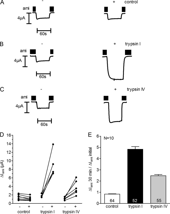
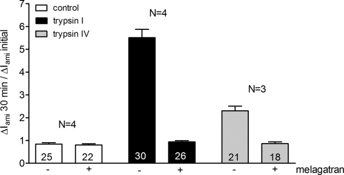
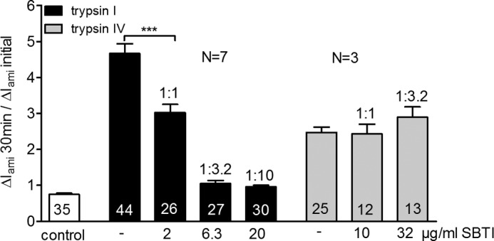
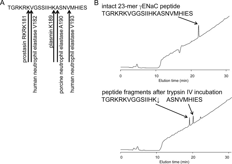
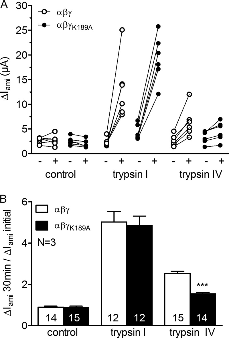
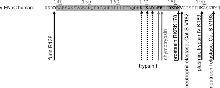
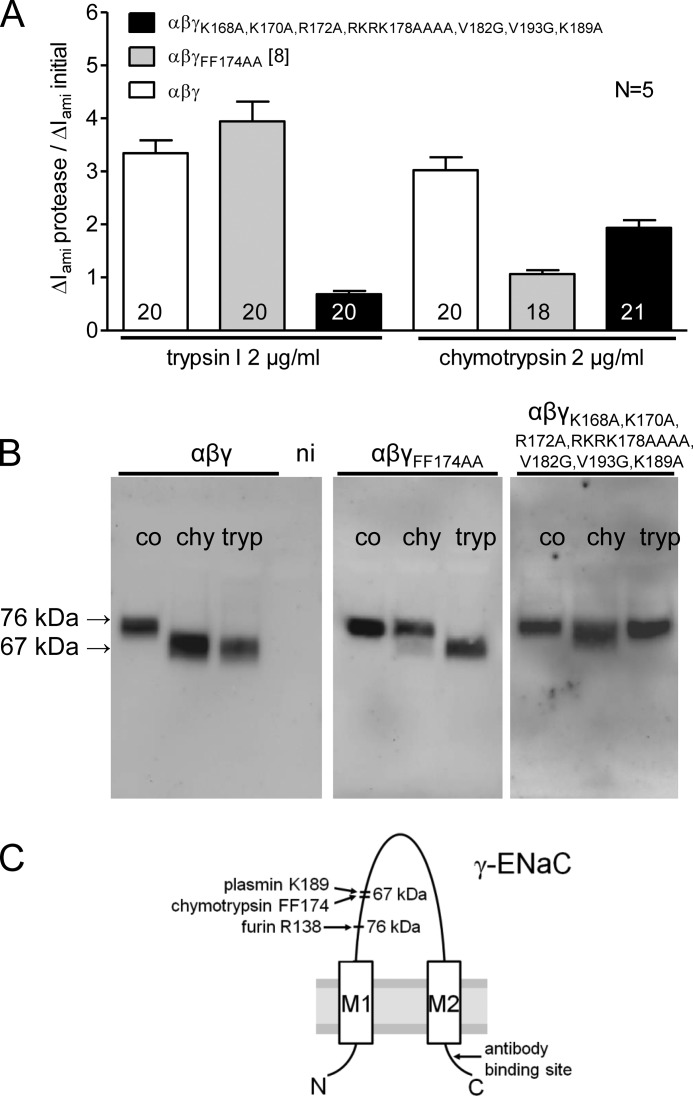
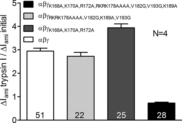
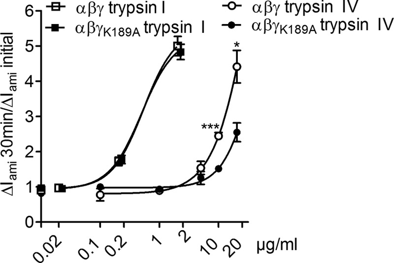
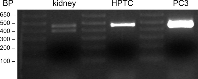
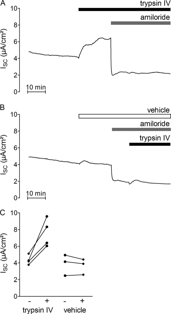
Proteolytic activation is a unique feature of the epithelial sodium channel (ENaC). However, the underlying molecular mechanisms and the physiologically relevant proteases remain to be identified. The serine protease trypsin I can activate ENaC in vitro but is unlikely to be the physiologically relevant activating protease in ENaC-expressing tissues in vivo. Herein, we investigated whether human trypsin IV, a form of trypsin that is co-expressed in several extrapancreatic epithelial cells with ENaC, can activate human ENaC. In Xenopus laevis oocytes, we monitored proteolytic activation of ENaC currents and the appearance of γENaC cleavage products at the cell surface. We demonstrated that trypsin IV and trypsin I can stimulate ENaC heterologously expressed in oocytes. ENaC cleavage and activation by trypsin IV but not by trypsin I required a critical cleavage site (Lys-189) in the extracellular domain of the γ-subunit. In contrast, channel activation by trypsin I was prevented by mutating three putative cleavage sites (Lys-168, Lys-170, and Arg-172) in addition to mutating previously described prostasin (RKRK(178)), plasmin (Lys-189), and neutrophil elastase (Val-182 and Val-193) sites. Moreover, we found that trypsin IV is expressed in human renal epithelial cells and can increase ENaC-mediated sodium transport in cultured human airway epithelial cells. Thus, trypsin IV may regulate ENaC function in epithelial tissues. Our results show, for the first time, that trypsin IV can stimulate ENaC and that trypsin IV and trypsin I activate ENaC by cleavage at distinct sites. The presence of distinct cleavage sites may be important for ENaC regulation by tissue-specific proteases.

Keywords: Epithelial Sodium Channel (ENaC); Ion Channel; Oocyte; Protease; Proteolytic Channel Activation; Serine Protease; Trypsin; Trypsin IV; Two-electrode Voltage Clamp.


Figures

FIGURE 1.
FIGURE 1.
Trypsin IV stimulates ENaC currents in X. laevis oocytes expressing human ENaC. Oocytes expressing human αβγENaC were preincubated for 30 min in protease-free solution (control) or in solution containing either trypsin I (2 μg/ml) or trypsin IV (10 μg/ml). Amiloride-sensitive whole-cell currents (ΔIami) were determined before (−) and after (+) incubation. A–C, six representative whole-cell current traces from one batch of oocytes are shown. Amiloride (ami) was present in the bath solution to specifically inhibit ENaC, as indicated by black bars. D, individual ΔIami values measured in one batch of oocytes. Data points obtained from an individual oocyte are connected by a line. E, summary of similar experiments as shown in D performed in 10 different batches of oocytes (n = 10). Columns represent the relative stimulatory effect of the incubation on ΔIami calculated as the ratio of ΔIami measured after 30-min incubation (ΔIami 30 min) to the initial ΔIamiIami initial) measured before incubation. The numbers inside the columns indicate the number of individual oocytes measured. Error bars, S.E.
FIGURE 2.
FIGURE 2.
The serine protease inhibitor melagatran prevents trypsin IV and trypsin I activation of ENaC. Oocytes expressing αβγENaC were incubated for 30 min in protease-free solution (control), in trypsin I (2 μg/ml), in trypsin IV (10 μg/ml), in melagatran (10 μm), in trypsin I (2 μg/ml) plus melagatran (10 μm), or in trypsin IV (10 μg/ml) plus melagatran (10 μm). Amiloride-sensitive whole-cell currents (ΔIami) were determined before and after incubation. Columns represent the relative effect of the incubation on ΔIami calculated as the ratio of ΔIami measured after 30-min incubation (ΔIami 30 min) to the initial ΔIamiIami initial) measured before incubation. The numbers inside the columns indicate the number of individual oocytes measured. N, number of different batches of oocytes used for the experiments. Error bars, S.E.
FIGURE 3.
FIGURE 3.
The polypeptide inhibitor SBTI prevents activation of ENaC by trypsin I but not trypsin IV. Oocytes expressing αβγENaC were incubated for 30 min in protease-free solution (control), in trypsin I (2 μg/ml), in trypsin IV (10 μg/ml), in trypsin I plus SBTI, or in trypsin IV plus SBTI. The ratio of protease to SBTI is indicated above the columns. Amiloride-sensitive whole-cell currents (ΔIami) were determined before and after incubation. Columns represent the relative effect of the incubation on ΔIami calculated as the ratio of ΔIami measured after 30-min incubation (ΔIami 30 min) to the initial ΔIamiIami initial) measured before incubation. The numbers inside the columns indicate the number of individual oocytes measured. N indicates the number of different batches of oocytes. ***, p < 0.001, unpaired t test. Error bars, S.E.
FIGURE 4.
FIGURE 4.
Trypsin IV cleaves within the extracellular domain of human γENaC. A, sequence of the 23-mer γENaC peptide showing putative cleavage sites for proteolytic ENaC activation. B, the 23-mer γENaC peptide (500 μm) was incubated with trypsin IV (50 μg/ml) for 30 min, and cleavage products were identified by HPLC and mass spectrometry.
FIGURE 5.
FIGURE 5.
Mutation of γK189A suppresses ENaC activation by trypsin IV, not trypsin I. Oocytes expressing αβγ (open symbols) or αβγK189AENaC (filled symbols) were incubated for 30 min in protease-free solution (control) or in a solution containing trypsin I (2 μg/ml) or trypsin IV (10 μg/ml). Amiloride-sensitive whole-cell currents (ΔIami) were determined before (−) and after (+) incubation. A, individual ΔIami values from a representative experiment using one batch of oocytes. Data points obtained from individual oocytes are connected by a line. B, summary of similar experiments as shown in A. Columns represent the relative effect of the incubation on ΔIami calculated as the ratio of ΔIami measured after 30-min incubation (ΔIami 30 min) to the initial ΔIamiIami initial) measured before incubation. Numbers inside the columns indicate the number of individual oocytes measured. N indicates the number of different batches of oocytes. ***, p < 0.001, unpaired t test. Error bars, S.E.
FIGURE 6.
FIGURE 6.
Sequence of human γENaC (amino acids 135–195). Amino acid sequence of human γENaC derived from the UniProt Database (UniProt number P51170). The putative cleavage sites for furin (Arg-138), trypsin I (Lys-168, Lys-170, and Arg-172), chymotrypsin (FF174), prostasin (RKRK178), human neutrophil elastase (Val-182 and Val-193), and plasmin (Lys-189) are indicated in boldface type and marked by arrows.
FIGURE 7.
FIGURE 7.
Mutating three putative trypsin I cleavage sites (Lys-168, Lys-170, and Arg-172) in addition to the putative prostasin, plasmin, and neutrophil elastase cleavage sites of ENaC prevents trypsin I activation of human ENaC. Continuous whole-cell current measurements were performed in oocytes expressing αβγ-, αβγFF174AA-, or αβγK168A,K170A,R172A,RKRK178AAAA,V182G,V193G,K189AENaC. ΔIami was determined before and after superfusing the oocytes with chymotrypsin (2 μg/ml) or trypsin I (2 μg/ml) (A). For biotinylation experiments, matched oocytes were preincubated for 30 min in protease-free solution (control; co) or in a solution containing either chymotrypsin (chy; 2 μg/ml) or trypsin I (tryp; 2 μg/ml). In parallel experiments, expression of biotinylated γENaC at the cell surface was analyzed by SDS-PAGE and followed by Western blot (B). A, columns represent relative effects of trypsin I or chymotrypsin on ΔIami calculated as the ratio of ΔIami after trypsin I or chymotrypsin superfusion (ΔIami protease) to the initial ΔIamiIami initial). The numbers inside the columns indicate the number of individual oocytes measured. N indicates the number of different batches of oocytes. B, representative Western blots from oocytes expressing αβγ, αβγFF174AA, or αβγK168A,K170A,R172A,RKRK178AAAA,V182G,V193G,K189AENaC. γENaC was detected with an antibody against the carboxyl terminus of human γENaC. In non-injected oocytes (ni), γENaC-specific signals were absent (data not shown). C, model of the γENaC subunit showing cleavage sites for proteolytic activation and the binding site of the antibody used. Error bars, S.E.
FIGURE 8.
FIGURE 8.
Combined mutation of the putative prostasin (RKRK178), plasmin (Lys-189), and neutrophil elastase (Val-182 and Val-193) cleavage sites or combined mutation of the three putative trypsin I cleavage sites (Lys-168, Lys-170, and Arg-172) of human γENaC does not reduce the stimulatory effect of trypsin I. Continuous whole-cell current measurements were performed in oocytes expressing αβγ-, αβγK168A,K170A,R172A-, αβγRKRK178AAAA,V182G,V193G,K189A-, or αβγK168A,K170A,R172A,RKRK178AAAA,V182G,V193G,K189AENaC. ΔIami was determined before and after superfusing the oocytes with trypsin I (2 μg/ml). The columns represent the relative effect of trypsin I on ΔIami calculated as the ratio of ΔIami after trypsin I superfusion (ΔIami trypsin I) to the initial ΔIamiIami initial). The numbers inside the columns indicate the number of individual oocytes measured. N indicates the number of different batches of oocytes. Error bars, S.E.
FIGURE 9.
FIGURE 9.
Effect of mutating a putative trypsin IV cleavage site (Lys-189) in γENaC on the concentration dependence of the stimulatory effect of trypsin IV and trypsin I on ENaC currents. Average concentration-response curves determined from experiments, as shown in Fig. 5. Oocytes expressing αβγ (open symbols) or αβγK189AENaC (filled symbols) were incubated for 30 min in solutions containing different concentrations of trypsin I or trypsin IV. Amiloride-sensitive whole-cell currents (ΔIami) were determined before (ΔIami initial) and after incubation (ΔIami 30 min). Each data point represents the average relative stimulatory effect of trypsin IV or trypsin I on ΔIami with 4–46 oocytes measured. Data were fitted using a sigmoidal dose response (variable slope). *, p < 0.05; ***, p < 0.001, unpaired t test. Error bars, S.E.
FIGURE 10.
FIGURE 10.
Expression of trypsinogen IV in human intact whole kidney and in HPTCs. PCR products for reverse-transcribed mRNA isolated from intact human kidney (left), from cultured HPTCs (middle), or from prostate cancer-derived human epithelial cells (PC3) (right) were obtained using the PCR primers and procedures outlined under “Experimental Procedures.” The sizes of the PCR products can be estimated from the base pair (BP) ladder standards shown.
FIGURE 11.
FIGURE 11.
Trypsin IV stimulates ENaC in human airway epithelial cells (H441). A and B, representative equivalent short circuit current (ISC) recordings from H441 distal airway epithelial cells. Trypsin IV (41 μg/ml) (A) or vehicle (0.9% NaCl) (B) was added to the apical bath solution of H441 cells as indicated by the horizontal bars. Amiloride (10 μm) was added apically to confirm that the stimulated ISC was mediated by ENaC. C, summary of results from similar experiments as shown in A and B. Data points represent individual ISC values obtained before and after (connected by a line) treatment with trypsin IV or vehicle.

Similar articles

Cited by

References

    1. Garty H., Palmer L. G. (1997) Epithelial sodium channels: function, structure, and regulation. Physiol. Rev. 77, 359–396 - PubMed
    1. Kellenberger S., Schild L. (2002) Epithelial sodium channel/degenerin family of ion channels: a variety of functions for a shared structure. Physiol. Rev. 82, 735–767 - PubMed
    1. Jasti J., Furukawa H., Gonzales E. B., Gouaux E. (2007) Structure of acid-sensing ion channel 1 at 1.9 Å resolution and low pH. Nature 449, 316–323 - PubMed
    1. Stockand J. D., Staruschenko A., Pochynyuk O., Booth R. E., Silverthorn D. U. (2008) Insight toward epithelial Na+ channel mechanism revealed by the acid-sensing ion channel 1 structure. IUBMB Life 60, 620–628 - PubMed
    1. Stewart A. P., Haerteis S., Diakov A., Korbmacher C., Edwardson J. M. (2011) Atomic force microscopy reveals the architecture of the epithelial sodium channel (ENaC). J. Biol. Chem. 286, 31944–31952 - PMC - PubMed

Publication types

LinkOut - more resources