

Mathematical modeling of sub-cellular asymmetry of fat-dachsous heterodimer for generation of planar cell polarity
- PMID: 24841507
- PMCID: PMC4026444
- DOI: 10.1371/journal.pone.0097641
Mathematical modeling of sub-cellular asymmetry of fat-dachsous heterodimer for generation of planar cell polarity
Abstract
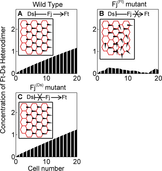
Planar Cell Polarity (PCP) is an evolutionarily conserved characteristic of animal tissues marked by coordinated polarization of cells or structures in the plane of a tissue. In insect wing epithelium, for instance, PCP is characterized by en masse orientation of hairs orthogonal to its apical-basal axis and pointing along the proximal-distal axis of the organ. Directional cue for PCP has been proposed to be generated by complex sets of interactions amongst three proteins - Fat (Ft), Dachsous (Ds) and Four-jointed (Fj). Ft and Ds are two atypical cadherins, which are phosphorylated by Fj, a Golgi kinase. Ft and Ds from adjacent cells bind heterophilically via their tandem cadherin repeats, and their binding affinities are regulated by Fj. Further, in the wing epithelium, sub-cellular levels of Ft-Ds heterodimers are seen to be elevated at the distal edges of individual cells, prefiguring their PCP. Mechanisms generating this sub-cellular asymmetry of Ft-Ds heterodimer in proximal and distal edges of cells, however, have not been resolved yet. Using a mathematical modeling approach, here we provide a framework for generation of this sub-cellular asymmetry of Ft-Ds heterodimer. First, we explain how the known interactions within Ft-Ds-Fj system translate into sub-cellular asymmetry of Ft-Ds heterodimer. Second, we show that this asymmetric localization of Ft-Ds heterodimer is lost when tissue-level gradient of Fj is flattened, or when phosphorylation of Ft by Fj is abolished, but not when tissue-level gradient of Ds is flattened or when phosphorylation of Ds is abrogated. Finally, we show that distal enrichment of Ds also amplifies Ft-Ds asymmetry. These observations reveal that gradient of Fj expression, phosphorylation of Ft by Fj and sub-cellular distal accumulation of Ds are three critical elements required for generating sub-cellular asymmetry of Ft-Ds heterodimer. Our model integrates the known experimental data and presents testable predictions for future studies.
Conflict of interest statement
Figures


 ) and that of (E–H) phosphorylated Ds (
 ) in cells over time. (A) and (E) show the randomized initial concentrations of
  and
and  , respectively.


 and
and  result in asymmetric localization of Ft-Ds heterodimer (µ = 1). Inset shows the enlarged view with inclined tops of the trapezoids. Data shown here is same as that shown in Figure 3H. (B) Higher contribution of sub-cellular asymmetry of
  augments the asymmetric localization of Ft-Ds heterodimer (steeper top of the trapezoids) in cells (µ = 10). (C) Higher contribution of
  sub-cellular asymmetry, however, results in diminished or even reversed asymmetry of Ft-Ds heterodimer in cells (µ = 0.1). Inset shows the enlarged view with flatter tops of the trapezoids indicating weak asymmetry of Ft-Ds heterodimer.
Similar articles
-
Expanding signaling-molecule wavefront model of cell polarization in the Drosophila wing primordium.PLoS Comput Biol. 2017 Jul 3;13(7):e1005610. doi: 10.1371/journal.pcbi.1005610. eCollection 2017 Jul. PLoS Comput Biol. 2017. PMID: 28671940 Free PMC article.
-
Planar cell polarity in the Drosophila eye is directed by graded Four-jointed and Dachsous expression.Development. 2004 Dec;131(24):6175-84. doi: 10.1242/dev.01550. Epub 2004 Nov 17. Development. 2004. PMID: 15548581
-
Microtubules provide directional information for core PCP function.Elife. 2014 Aug 14;3:e02893. doi: 10.7554/eLife.02893. Elife. 2014. PMID: 25124458 Free PMC article.
-
Conservation of Planar Polarity Pathway Function Across the Animal Kingdom.Annu Rev Genet. 2015;49:529-51. doi: 10.1146/annurev-genet-112414-055224. Epub 2015 Sep 10. Annu Rev Genet. 2015. PMID: 26360326 Review.
-
Regulation of PCP by the Fat signaling pathway.Genes Dev. 2013 Oct 15;27(20):2207-20. doi: 10.1101/gad.228098.113. Genes Dev. 2013. PMID: 24142873 Free PMC article. Review.
Cited by
-
Planar cell polarity of the kidney.Exp Cell Res. 2016 May 1;343(2):258-266. doi: 10.1016/j.yexcr.2014.11.003. Epub 2014 Nov 13. Exp Cell Res. 2016. PMID: 25445789 Free PMC article. Review.
-
Jagged mediates differences in normal and tumor angiogenesis by affecting tip-stalk fate decision.Proc Natl Acad Sci U S A. 2015 Jul 21;112(29):E3836-44. doi: 10.1073/pnas.1511814112. Epub 2015 Jul 7. Proc Natl Acad Sci U S A. 2015. PMID: 26153421 Free PMC article.
-
Expanding signaling-molecule wavefront model of cell polarization in the Drosophila wing primordium.PLoS Comput Biol. 2017 Jul 3;13(7):e1005610. doi: 10.1371/journal.pcbi.1005610. eCollection 2017 Jul. PLoS Comput Biol. 2017. PMID: 28671940 Free PMC article.
-
Distinct mechanisms of planar polarization by the core and Fat-Dachsous planar polarity pathways in the Drosophila wing.Cell Rep. 2022 Sep 27;40(13):111419. doi: 10.1016/j.celrep.2022.111419. Cell Rep. 2022. PMID: 36170824 Free PMC article.
-
Heterophilic cell-cell adhesion of atypical cadherins Fat and Dachsous regulate epithelial cell size dynamics during Drosophila thorax morphogenesis.Mol Biol Cell. 2020 Mar 19;31(7):546-560. doi: 10.1091/mbc.E19-08-0468. Epub 2019 Dec 26. Mol Biol Cell. 2020. PMID: 31877063 Free PMC article.
References
Publication types
MeSH terms
Substances
LinkOut - more resources
Full Text Sources
Other Literature Sources
Molecular Biology Databases
Research Materials
