. 2014 Sep;357(3):719-30.
doi: 10.1007/s00441-014-1879-8. Epub 2014 May 20.

Activation of spleen cells by ArtinM may account for its immunomodulatory properties

Affiliations

Activation of spleen cells by ArtinM may account for its immunomodulatory properties

Thiago Aparecido da Silva et al. Cell Tissue Res. 2014 Sep.

Abstract

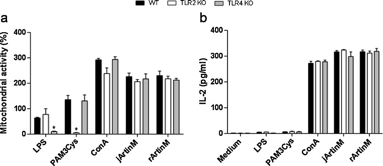
ArtinM is a D-mannose-binding lectin extracted from Artocarpus heterophyllus that promotes interleukin-12 production by macrophages and dendritic cells. This property is considered responsible for T helper 1 immunity induced in vivo after ArtinM administration. In this study, we investigated the effect of native (jArtinM) and recombinant (rArtinM) forms of lectin on murine spleen cells and isolated T lymphocytes. We found that ArtinM binds to the surface of spleen cells. This interaction, which was blocked by D-mannose, induced cell activation, as manifested by increased mitochondrial activity, interleukin-2 production, and cell proliferation. We verified that a 30-times higher concentration of rArtinM was required to trigger optimal activation of spleen cells compared with that needed with jArtinM, although these proteins have identical sugar recognition properties and use the same signaling molecules to trigger cell activation. Because the distinction between native and recombinant is restricted to their tertiary structure (tetrameric and monomeric, respectively), we postulated that the multi-valence of jArtinM accounts for its superiority in promoting clustering of cell surface glycoreceptors and activation. The jArtinM and rArtinM activation effect exerted on spleen cells was reproduced on purified CD4(+) T cells. Our results suggest that ArtinM interaction with T cells leads to responses that may act in concert with the interleukin-12 produced by antigen-presenting cells to modulate immunity toward the T helper 1 axis. Further studies are necessary to dissect ArtinM/T-cell interactions to more fully understand the immunomodulation induced by carbohydrate recognition.


Figures

Fig. 1
Fig. 1
ArtinM binds to the surface of spleen cells through carbohydrate recognition. Biotinylated jArtinM (20 μg/mL) was incubated with the indicated concentrations of D-mannose (cg) or D-galactose (hl). The resulting mixtures were added to a suspension (1.5 × 106 cells/mL) of fixed spleen cells, which were obtained from BALB/c mice. ArtinM binding was revealed by reaction with streptavidin-FITC (5 μg/mL). Streptavidin-FITC alone was used as a negative control (a). The cells were washed and analyzed for fluorescence intensity by flow cytometry. Figures show the percentage of positive cells for ArtinM binding for each experimental condition (bl). The results are expressed as means ± SEM and represent 3 independent experiments
Fig. 2
Fig. 2
ArtinM stimulates mitochondrial activity of spleen cells in a dose-dependent manner. Murine spleen cells (1.5 × 106 cells/mL) from BALB/c were distributed in 96-well microplates and incubated at 37 °C in a humidified atmosphere of 5 % CO2 and stimulated with jArtinM (a) or rArtinM (b) in concentrations of 0.14–156 or 0.56–625 nM, respectively. Non-stimulated spleen cells were used as negative controls. After 12, 24, 48, and 72 h of incubation, 3-(4,5-dimethyl-thiazol-2-yl)-2,5-diphenyltetrazolium bromide (MTT) was added to the culture medium; MTT reduction to insoluble purple formazan dye crystals was detected via absorbance reading at 570 nm. Mitochondrial activity was expressed as absorbance variation (in percentages) in relation to the negative control. Stimulation with Concanavalin A (49 nM) was used as a positive control, which provided the following absorbance variations: 114.9 ± 4.7 (12 h), 240.6 ± 40.58 (24 h), 852.7 ± 22.41 (48 h), and 704.6 ± 15.3 (72 h). The results represent 3 independent experiments and are expressed as means ± SEM
Fig. 3
Fig. 3
ArtinM stimulates interleukin-2 (IL-2) production by spleen cells in a dose-dependent manner. Spleen cells (1.5 × 106 cells/mL) from BALB/c were distributed in 96-well microplates and incubated at 37 °C in a humidified atmosphere of 5 % CO2 and stimulated with jArtinM (a) or rArtinM (b) in concentrations of 0.14–156 or 0.56–625 nM, respectively. After 12, 24, 48, and 72 h of incubation, the cell culture supernatants were analyzed for IL-2 levels with enzyme-linked immunosorbent assay. Stimulation with Concanavalin A (49 nM), used as a positive control, provided the following IL-2 levels: 465.60 ± 4.49 (12 h), 495.8 ± 7.06 (24 h), 421.6 ± 4.37 (48 h), and 431.3 ± 6.73 (72 h). Non-stimulated spleen cells, used as negative controls (medium), provided the following IL-2 levels: 10.11 ± 2.04 (12 h), 10.24 ± 2.16 (24 h), 18.19 ± 3.29 (48 h), and 31.59 ± 4.49 (72 h). The results represent 3 independent experiments and are expressed as means ± SEM. *p < 0.05 compared to the medium for all incubation periods
Fig. 4
Fig. 4
Spleen cell proliferation induced by ArtinM. Spleen cells (1.5 × 106 cells/mL) from BALB/c were distributed in a 96-well microplate and incubated for 48 h at 37 °C in a humidified atmosphere with 5 % CO2 and stimulated with the indicated concentrations of jArtinM or rArtinM. Medium alone and Concanavalin A (ConA) (24.5 nM) were used as negative and positive controls, respectively. After 36 h of stimulation, [3H]-thymidine (0.5 μCi/well) was added and measured for incorporation (cpm) after 12 h of incubation. The results are expressed as means ± SEM. *Significant differences with p < 0.05 in relation to medium values
Fig. 5
Fig. 5
Spleen cell activation by ArtinM depends on carbohydrate recognition. Murine spleen cells (1.5 × 106 cells/mL) obtained from BALB/c were distributed in a 96-well microplate and incubated at 37 °C in a humidified atmosphere of 5 % CO2. The cells were stimulated with jArtinM (2.25 nM) or rArtinM (78.00 nM) and were previously incubated or not with 50 mM D-mannose or D-galactose. Phorbol myristate acetate (PMA; 81 nM) plus ionomycin (1 μM) was used as a positive control stimulus. After 48 h of stimulation, mitochondrial activity (a) and IL-2 production (b) were determined. The results are expressed as % of inhibition, obtained by relating the measurements in the presence and absence of sugar for each stimulus. The results represent 3 independent experiments
Fig. 6
Fig. 6
TLR2 or TLR4 does not mediate spleen cell activation by ArtinM. Murine spleen cells (1.5 × 106 cells/mL) from C57BL/6 (WT), TLR2 KO, and TLR4 KO mice were plated onto 96-well microplates, incubated at 37 °C and 5 % CO2 in a humidified incubator, and stimulated for 24 h with 9 nM jArtinM or 312 nM rArtinM. Pam3Cys (TLR2 agonist, 1.5 μg/mL), LPS (TLR4 agonist, 1 μg/mL), or Concanavalin A (ConA) (49 nM), were used as positive controls for stimulation, whereas the medium alone was used as negative control. Next, the cells were analyzed for mitochondrial activity (MTT assay) (a) and IL-2 production (ELISA method) (b). Results are expressed as means ± SEM. *Differences in response from that of WT cells with p < 0.05 was considered as significant
Fig. 7
Fig. 7
Effect of inhibitor agents of molecules accounting for cell signaling pathways on jArtinM- or rArtinM-induced activation of spleen cells. PD98059 [p42/44 mitogen-associated protein kinase (MAPK) inhibitor; 20 μM], genistein (protein tyrosine kinase inhibitor; 20 μg/mL), SB202190 (p38 MAPK inhibitor; 20 μM), SP600125 (c-Jun N-terminal kinase inhibitor; 25 μM), H-7 (protein kinase C inhibitor; 20 μM), or medium alone (absence of inhibitor) were used to pretreat, for 210 min, murine spleen cells (1.5 × 106/mL) that were distributed in 96-well plates and maintained at 37 °C in a humidified atmosphere with 5 % CO2. After pretreatment, cells were stimulated with jArtinM (2.25 nM) or rArtinM (78 nM) for 48 h. PMA (81 nM) plus ionomycin (1 μM) or Concanavalin A (ConA) (49 nM) were positive controls for cell stimulation, whereas medium alone provided the negative control. The cells were analyzed for mitochondrial activity via MTT assay (a) and IL-2 production via ELISA (b). Measurements are expressed as means ± SEM, and the inhibition determined by each pharmacological agent was calculated by the ratio between the values obtained in the presence of a certain inhibitor and in its absence, represented by percentages
Fig. 8
Fig. 8
ArtinM binds to the surface of T lymphocytes. Spleen cells (1.5 × 106/ml) were fixed and incubated for 40 min with anti-CD3 PE-Cy5 antibody (4 μg/mL) (b, d). After washing, the cells were incubated for 40 min with biotinylated ArtinM (15 μg/mL) (c, d). ArtinM binding was revealed by reaction for 40 min with streptavidin-FITC (5 μg/mL). Streptavidin-FITC and Isotype PE-Cy5 were used as a negative control (a). Cell fluorescence was analyzed with flow cytometry. Dot plots represent the percentage of positive cells obtained by biotinyl-ArtinM + strp/FITC or anti-CD3 PE-Cy5. The results are expressed as means ± SEM
Fig. 9
Fig. 9
jArtinM and rArtinM enhances IL-2 secretion in CD4+ T cells. Purified CD4+ T cells (1 × 106 cells/mL) from a spleen cell suspension of BALB/c mice were distributed in 96-well microplates and maintained at 37 °C in a humidified atmosphere with 5 % CO2. The CD4+ T cells were stimulated for 48 h with jArtinM (18, 36, or 78 nM) or rArtinM (78, 156, or 312 nM). Concanavalin A (49 nM) was used as a positive control of stimulation. Culture supernatants were analyzed for IL-2 production using ELISA, and the results are expressed as means ± SEM. *Significant differences when p < 0.05 compared with medium values
Fig. 10
Fig. 10
CD3 receptor as a glycotarget of ArtinM. (ac) Purified CD4+ T cells (1.5 × 106/mL) were fixed and incubated for 40 min with ArtinM (25 μg/mL). After washing, the cells were incubated for 40 min with an anti-CD3 PE-Cy5 antibody (10 μg/mL) and analyzed using flow cytometry. The histograms represent the percentage of cells positive for staining with the anti-CD3 PE-Cy5 antibody after preincubation with (b) or without (a) ArtinM. d Isolated CD4+ T cells (1 × 106/mL) were incubated with the anti-CD3 antibody (5 μg/mL) or an anti-CD28 antibody (5 μg/mL) for 40 min and then stimulated with ArtinM (78 nM) for 48 h. IL-2 concentration in the culture supernatants was quantified by means of ELISA, and the results are expressed as mean ± SEM; *p < 0.05 compared to the cells that were preincubated with vehicle (culture medium) and then stimulated with ArtinM

Similar articles

Cited by

References

    1. Baba M, Yong Ma B, Nonaka M, Matsuishi Y, Hirano M, Nakamura N, Kawasaki N, Kawasaki T. Glycosylation-dependent interaction of Jacalin with CD45 induces T lymphocyte activation and Th1/Th2 cytokine secretion. J Leukoc Biol. 2007;81:1002–1011. doi: 10.1189/jlb.1106660. - DOI - PubMed
    1. Benoist H, Culerrier R, Poiroux G, Segui B, Jauneau A, Van Damme EJ, Peumans WJ, Barre A, Rouge P. Two structurally identical mannose-specific jacalin-related lectins display different effects on human T lymphocyte activation and cell death. J Leukoc Biol. 2009;86:103–114. doi: 10.1189/jlb.0708434. - DOI - PubMed
    1. Cardoso MR, Mota CM, Ribeiro DP, Santiago FM, Carvalho JV, Araujo EC, Silva NM, Mineo TW, Roque-Barreira MC, Mineo JR, Silva DA (2011) ArtinM, a D-mannose-binding lectin from Artocarpus integrifolia, plays a potent adjuvant and immuno stimulatory role in immunization against Neospora caninum. Vaccine 29:9183–9193 - PubMed
    1. Coltri KC, Oliveira LL, Pinzan CF, Vendruscolo PE, Martinez R, Goldman MH, Panunto-Castelo A, Roque-Barreira MC. Therapeutic administration of KM + lectin protects mice against Paracoccidioides brasiliensis infection via interleukin-12 production in a toll-like receptor 2-dependent mechanism. Am J Pathol. 2008;173:423–432. doi: 10.2353/ajpath.2008.080126. - DOI - PMC - PubMed
    1. Coltri KC, Oliveira LL, Ruas LP, Vendruscolo PE, Goldman MH, Panunto-Castelo A, Roque-Barreira MC. Protection against Paracoccidioides brasiliensis infection conferred by the prophylactic administration of native and recombinant ArtinM. Med Mycol. 2010;48:792–799. doi: 10.3109/13693780903501671. - DOI - PubMed

Publication types

MeSH terms

LinkOut - more resources