. 2014 May 19;9(5):e97835.
doi: 10.1371/journal.pone.0097835. eCollection 2014.

Apple derived cellulose scaffolds for 3D mammalian cell culture

Affiliations

Apple derived cellulose scaffolds for 3D mammalian cell culture

Daniel J Modulevsky et al. PLoS One. .

Abstract

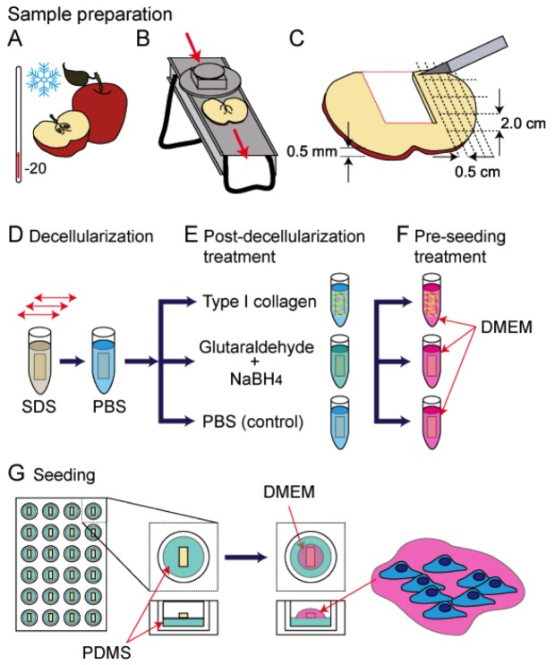
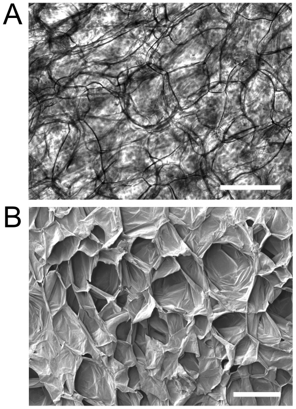
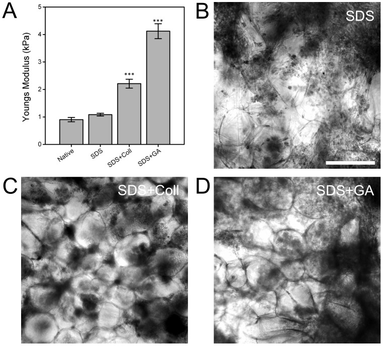
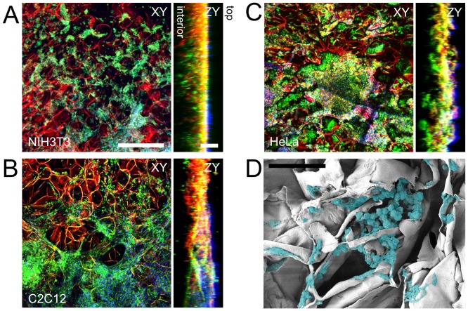
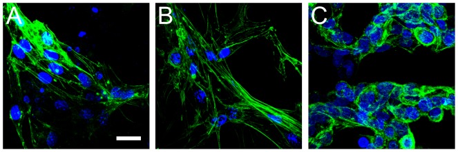
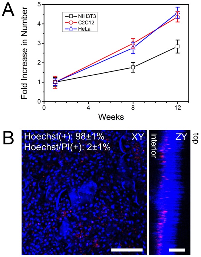
There are numerous approaches for producing natural and synthetic 3D scaffolds that support the proliferation of mammalian cells. 3D scaffolds better represent the natural cellular microenvironment and have many potential applications in vitro and in vivo. Here, we demonstrate that 3D cellulose scaffolds produced by decellularizing apple hypanthium tissue can be employed for in vitro 3D culture of NIH3T3 fibroblasts, mouse C2C12 muscle myoblasts and human HeLa epithelial cells. We show that these cells can adhere, invade and proliferate in the cellulose scaffolds. In addition, biochemical functionalization or chemical cross-linking can be employed to control the surface biochemistry and/or mechanical properties of the scaffold. The cells retain high viability even after 12 continuous weeks of culture and can achieve cell densities comparable with other natural and synthetic scaffold materials. Apple derived cellulose scaffolds are easily produced, inexpensive and originate from a renewable source. Taken together, these results demonstrate that naturally derived cellulose scaffolds offer a complementary approach to existing techniques for the in vitro culture of mammalian cells in a 3D environment.


Conflict of interest statement

Competing Interests: Andrew Pelling is a PLOS ONE editorial board member and this does not alter the authors' adherence to all the PLOS ONE policies on sharing data and materials.

Figures

Figure 1
Figure 1. A cartoon schematic representing the apple tissue decellularization and mammalian cell seeding protocol used in this study.
A) McIntosh Red apples were exposed to −20°C temperatures for a max duration of 5 minutes, to increase the firmness of the outer apple hypanthium tissue. B) Uniform 1.2±0.1 mm thick slices of the apples were obtained using a mandolin slicer. Slices containing any of the ovary core of the apple were removed. C) The apple slices were cut into uniform 2.0 by 0.5 cm segments that were placed in individual microcentrifuge tubes. D) A 0.5% SDS solution was added to the microcentrifuge tubes and placed on a shaker for 12 hours at room temperature. The scaffolds were then rinsed repeatedly with PBS and allowed to incubate in a PBS solution with 1% streptomycin/penicillin and 1% amphotericin B for 6 hours at room temperature. E) The scaffolds were then coated with Type 1 collagen, chemically cross linked with glutaraldehyde or incubated in PBS. F) All the samples were then incubated in mammalian cell culture medium (DMEM) for 12 hours in a standard tissue culture incubator maintained at 37°C and 5% CO2. G) The scaffolds were placed in PDMS coated 24 well plates and a 40 µL cell suspension was placed on each. After 6 hours in the incubator the wells were filled with DMEM and cells cultured for up to 12 weeks.
Figure 2
Figure 2. Decellularized cellulose scaffolds.
A) Phase contrast image of cellulose cell wall structure in a decellularized apple tissue sample. The dark lines correspond to distinct cellulose structures which form a three dimensional matrix. B) SEM image of a similar cellulose scaffold revealing its three dimensional nature and large cavities. Scale bar = 200 µm.
Figure 3
Figure 3. The mechanical properties of functionalized cellulose 3D scaffolds and C2C12 myoblast cultured within the 3D cellulose scaffolds.
A) The local mechanical elasticity of native tissue, decellularized (SDS), collagen functionalized (SDS+Coll) and glutaraldehyde (SDS+GA) cross-linked cellulose scaffolds. The native tissue and unmodified scaffolds do not display any significant difference in mechanical properties. Both the collagen functionalized and chemically cross-linked scaffolds displayed a significant increase in elasticity compared to the DMEM scaffolds (*** = p<0.001). The (B) decellularized (SDS), (C) Collagen functionalized (SDS+Coll) and (D) glutaraldheyde cross-linked (SDS+GA) scaffolds all support the growth of C2C12 cells. Phase contrast images of C2C12 cells after two weeks of growth reveal the presence of cell colonies. Scale bar = 200 µm.
Figure 4
Figure 4. Fixed and stained NIH3T3, C2C12 and HeLa cells cultured on 3D cellulose scaffolds.
Specific fluorescent staining of (A) NIH3T3, (B) C2C12 and (C) HeLa mammalian cells within the cellulose scaffolds and subsequent laser scanning confocal microscopy reveals the cellulose structure (red), mammalian cell membranes (green) and nuclei (blue). Cells were cultured in these scaffolds for four weeks prior to staining and imaging. Confocal volumes were acquired and projected in the XY and ZY plane. The ZY orthogonal views demonstrate the depth of cell proliferation within the cellulose scaffold. The top and bottom surfaces of the scaffold are indicated. Scale bars: XY = 300 µm, ZY = 100 µm. D) SEM image of a cellulose scaffold cross section after being seeded with C2C12 cells that were allowed to proliferate for four weeks. The cells were digitally colourized in order to increase contrast between the cells and cellulose structure (Scale bar: 50 µm)
Figure 5
Figure 5. Fixed and stained images of cells actin cytoskeleton cultured within the 3D cellulose scaffold.
A) NIH3T3, B) C2C12 and C) HeLa cells were cultured onto the cellulose scaffolds for 2 weeks prior to stained for actin (green) and cell nuclei (blue). (. NIH3T3 and C2C12 cells display characteristic actin stress fibres found in cultured cells. HeLa cells also display characteristic actin structures including fewer prominent stress fibres and a large amount of cortical actin localization. Scale bar = 25 µm and applies to all.
Figure 6
Figure 6. Cell proliferation and viability over time.
A) NIH3T3, C2C12 and HeLa cells were cultured individually in cellulose n = 3 scaffolds for 1, 8 and 2 weeks and then imaged with confocal microscopy after being stained with Hoechst 33342. Cells were counted at each time point and an increase in cell population is clearly observed. B) After 12 weeks of culture, C2C12 cells were fixed and stained with Hoechst 33342 (blue: viable cells) and Propidium iodide (PI) (red: apoptotic/necrotic cells). Confocal volumes were acquired and projected in the XY and ZY plane and reveal that cells have proliferated throughout the structure during the 12-week culture. The cells that are apoptotic/necrotic are found in deeper regions of the scaffold. The top and bottom surfaces of the scaffold are indicated. The number of live (Hoechst(+)) and dead (Hoechst/PI(+)) cells were counted and it was found that ∼98% of the cells within the scaffold are viable. Data is shown for C2C12 cells, but is similar for NIH3T3 and HeLa cells (data not shown). Scale bar: B = 200 µm for XY and 100 µm for ZY.

Similar articles

Cited by

References

    1. Yamada KM, Cukierman E (2007) Modeling tissue morphogenesis and cancer in 3D. Cell 130: 601–610 10.1016/j.cell.2007.08.006 - DOI - PubMed
    1. Derda R, Laromaine A, Mammoto A, Tang SKY, Mammoto T, et al. (2009) Paper-supported 3D cell culture for tissue-based bioassays. PNAS 106: 18457–18462 10.1073/pnas.0910666106 - DOI - PMC - PubMed
    1. McBane JE, Sharifpoor S, Cai K, Labow RS, Santerre JP (2011) Biodegradation and in vivo biocompatibility of a degradable, polar/hydrophobic/ionic polyurethane for tissue engineering applications. Biomaterials 32: 6034–6044 10.1016/j.biomaterials.2011.04.048 - DOI - PubMed
    1. Haycock JW (2011) 3D Cell Culture: A Review of Current Approaches and Techniques. Methods Mol Biol 695: 1–15 10.1007/978-1-60761-984-0 - DOI - PubMed
    1. Puschmann TB, Zandén C, De Pablo Y, Kirchhoff F, Pekna M, et al. (2013) Bioactive 3D cell culture system minimizes cellular stress and maintains the in vivo-like morphological complexity of astroglial cells. Glia 61: 432–440 10.1002/glia.22446 - DOI - PubMed

Publication types