. 2014 Aug;34(15):2822-32.
doi: 10.1128/MCB.00206-14. Epub 2014 May 19.

Regulated proteolysis of NOTCH2 and NOTCH3 receptors by ADAM10 and presenilins

Affiliations

Regulated proteolysis of NOTCH2 and NOTCH3 receptors by ADAM10 and presenilins

Arjan J Groot et al. Mol Cell Biol. 2014 Aug.

Abstract

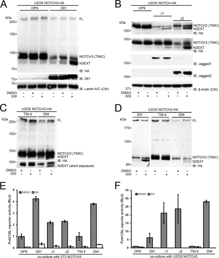
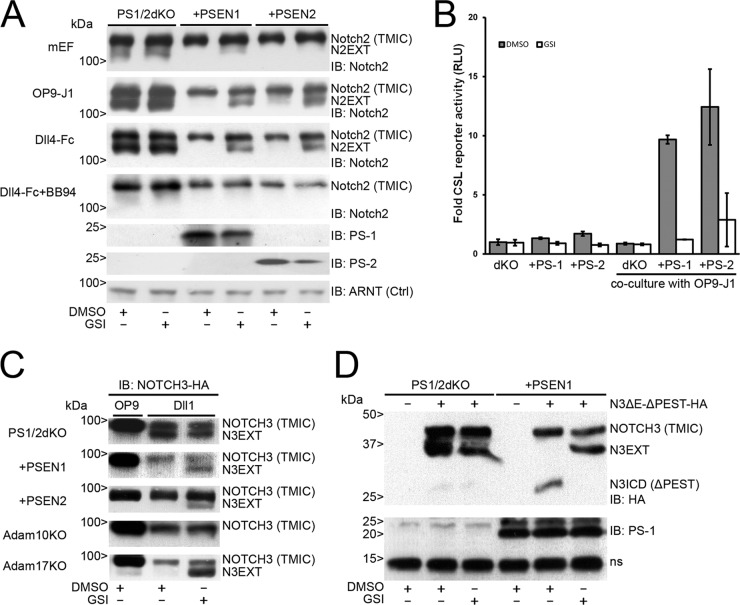
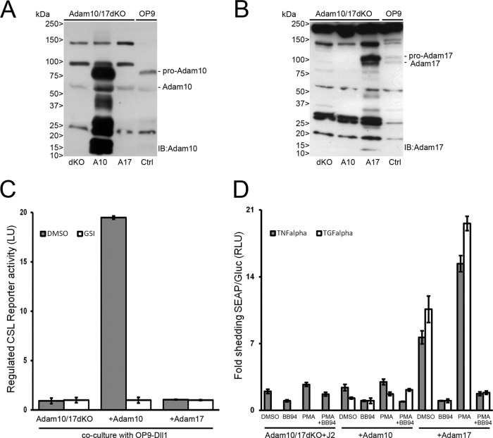
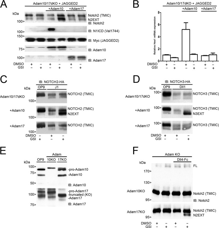
In mammals, there are four NOTCH receptors and five Delta-Jagged-type ligands regulating many aspects of embryonic development and adult tissue homeostasis. NOTCH proteins are type I transmembrane receptors that interact with ligands on adjacent cells and are activated by regulated intramembrane proteolysis (RIP). The activation mechanism of NOTCH1 receptors upon ligand binding is well understood and requires cleavage by ADAM10 metalloproteases prior to intramembranous cleavage by γ-secretase. How the other human NOTCH receptor homologues are activated upon ligand binding is not known. Here, we dissect the proteolytic activation mechanism of the NOTCH2 and NOTCH3 receptors. We show that NOTCH2 and NOTCH3 signaling can be triggered by both Delta-Jagged-type ligands and requires ADAM10 and presenilin-1 or -2. Importantly, we did not find any role for the highly related ADAM17/TACE (tumor necrosis factor alpha-converting enzyme) protease in ligand-induced NOTCH2 or NOTCH3 signaling. These results demonstrate that canonical ligand-induced proteolysis of the NOTCH1, -2, and -3 receptors strictly depends on consecutive cleavage of these receptors by ADAM10 and the presenilin-containing γ-secretase complex, leading to transcriptional activation.


Figures

FIG 1
FIG 1
Human NOTCH2 and NOTCH3 proteolytic processing and transcriptional activation are triggered by DSL ligands. (A) Coculture experiment of U2OS NOTCH3-HA cells and OP9 parental or Dll1-expressing cells in the absence or presence of GSI. DMSO was used as a vehicle control. (Top panel) HA immunoblotting of cell lysates shows expression of both the full-length (FL) NOTCH3 precursor and mature, S1-processed, NOTCH3 TMIC. Dll1 stimulation leads to activation and diminished levels of NOTCH3 TMIC and accumulation of N3EXT in the presence of GSI. (Middle panel) Dll1 immunoblotting confirms expression of Dll1, which is absent in OP9 parental cells. (Bottom panel) Lamin A/C immunoblotting serves as a loading control (Ctrl). (B) Coculture experiment of U2OS NOTCH2-HA- and NOTCH3-HA-expressing cells with OP9 parental or Jagged1 (J1)- or Jagged2 (J2)-overexpressing cells in the absence or presence of GSI. (Top two panels) Stimulation by J1 and J2 leads to activation and diminished levels of NOTCH2 and NOTCH3 TMIC. N2EXT and N3EXT accumulate in the presence of GSI. (Middle two panels) Immunoblotting confirms expression of J1 and J2, which are absent in OP9 parental cells. (Bottom panel) β-Actin immunoblotting serves as a loading control (Ctrl). (C) Coculture experiment of U2OS NOTCH3-HA cells on top of TSt-4 parental or Dll4-overexpressing cells in the absence or presence of GSI. HA immunoblotting shows that Dll4 stimulation leads to NOTCH3 receptor proteolysis diminished TMIC expression and GSI-dependent accumulation of N3EXT (short exposure). (D) Coculture experiment of U2OS NOTCH2-HA-expressing cells with Dll1 or TSt-4 parental and TSt-4–Dll4-overexpressing cells in the absence or presence of GSI. HA immunoblotting of cell lysates shows expression of both the full-length (FL) NOTCH2 precursor and S1-processed NOTCH2 TMIC. Dll1 (left) and Dll4 (right) were able to activate NOTCH2 proteolytic processing, as accumulation of N2EXT fragments could be visualized by addition of GSI. Molecular mass marker proteins are indicated. (E and F) Dual NOTCH (CSL) luciferase reporter gene activity, corrected for Renilla luciferase expression, in either 3T3 cells expressing NOTCH3 (E) or U2OS cells expressing NOTCH2 (F). In response to coculture with ligand-expressing cells, an increased transcriptional activation was observed in comparison to coculture without ligand (OP9/TSt-4), which was completely blocked by GSI treatment. NOTCH-dependent signals were in parental cells arbitrarily set to 1. Measurements correspond to at least two experiments in triplicate and are displayed as relative light units (RLU). Error bars represent means ± standard deviations.
FIG 2
FIG 2
Notch2 and NOTCH3 proteolytic processing is regulated by presenilin-1 or -2. (A) Immunoblots showing expression of endogenous Notch2 in lysates from mouse embryonic fibroblasts (mEF) with double knockout for presenilin-1 and -2 (PS1/2dKO) or reconstituted with presenilin-1 (PSEN1) or 2 (PSEN2) (top panel), in coculture with OP9-J1 cells (second panel) or with coated Dll4-Fc molecules (third panel) in the absence or presence of GSI and GSI/BB94 (fourth panel). Immunoblots for presenilin-1 and -2 proteins (PS-1 and PS-2) show reconstitution of the respective presenilin, as indicated. ARNT (aryl hydrocarbon receptor nuclear translocator) served as a loading control (Ctrl). PS1/2dKO cells were unable to S3 process Notch2, indicated by accumulated N2EXT fragments after stimulation by either J1 or recombinant Dll4-Fc molecules. In the cells with PSEN1 or PSEN2, N2EXT fragments were observed only in the presence of GSI. Treatment with BB94 abrogates formation of N2EXT, indicating that the formation of N2EXT is dependent on metalloproteases. (B) Dual Notch (CSL) luciferase reporter gene activity, corrected for Renilla luciferase expression, in either PS1/2dKO cells or cells reconstituted with presenilin-1 (PS-1) or -2 (PS-2). In response to coculture with Jagged1-expressing cells, a γ-secretase-dependent increased transcriptional activation was observed only in cells reconstituted with either PS-1 or PS-2 and not in PS1/2dKO cells. All values were normalized to DMSO-treated PS1/2dKO cells and arbitrarily set to 1. Measurements correspond to at least two experiments in triplicate and are displayed as relative light units (RLU). Error bars represent means ± standard deviations. (C) Coculture experiment of NOTCH3-HA-expressing PS1/2dKO cells reconstituted with either PSEN1 or PSEN2, Adam10KO, and Adam17KO cells with Dll1-expressing cells compared to OP9 parental cells. HA immunoblots show diminished levels of TMIC after stimulation with Dll1. (Top three panels) N3EXT fragments accumulate in the absence of presenilin proteins, whereas in the presence of either PSEN1 or PSEN2, N3EXT accumulation was observed only when treated with GSI. (Bottom two panels) N3EXT fragments were absent in Adam10KO cells but not in Adam17KO cells after stimulation by Dll1 in the presence of GSI. (D) (Top) HA immunoblotting of cell lysates of nontransfected PS1/2dKO and PSEN1 reconstituted cells or transfected with an EGF repeat and PEST domain NOTCH3-HA deletion construct (hN3ΔE-ΔPEST) in the absence or presence of GSI. N3EXT accumulation was observed in hNOTCH3ΔE-ΔPEST-expressing PS1/2dKO cells without ligand stimulation. In PS1/2dKO cells reconstituted with PSEN1, N3EXT accumulation could be observed only in the presence of GSI. Furthermore, truncated N3ICD fragments were observed in PS1/2dKO cells reconstituted with PSEN1, which were absent in the presence of GSI. (Bottom) Immunoblot showing PS-1 expression with a nonspecific band (ns). Molecular masses are indicated.
FIG 3
FIG 3
Characterization and functional analysis of Adam10/17dKO or Adam10/Adam17-reconstituted MEFs. (A and B) Immunoblotting with Adam10 and Adam17 antibodies on lysates from Adam10/17dKO MEF cells reconstituted with either Adam10 (A10) or Adam17 (A17). Pro- and mature Adam10 proteins could be detected only in Adam10-reconstituted Adam10/17dKO or OP9 cells (Ctrl), whereas Adam17 pro- and mature proteins were detected only in Adam17-reconstituted Adam10/17dKO or Ctrl cells. Molecular masses are indicated. (C) Firefly luciferase transcriptional Notch CSL reporter activity in Adam10/17dKO cells, reconstituted with either Adam10 or Adam17, cocultured with OP9-Dll1 cells. Regulated Notch reporter activity in light units (LU) could be measured only in cells expressing Adam10, which could be inhibited by GSI. Measurements were arbitrarily set to GSI values and correspond to at least two experiments in triplicate. Error bars represent means ± standard deviations. (D) AP-TNF-α- and AP-TGF-α-transfected Adam10/17dKO-J2 MEF cells reconstituted with either Adam10 or Adam17 were stimulated with DMSO (vehicle), BB94 (5 μM), PMA (100 ng/ml), or both BB94 and PMA. Gaussia luciferase excretion, unaffected by any of the stimuli, was used as an internal control. Medium was analyzed 1 h after stimulation for the presence of shed AP-TNF-α and AP-TGF-α molecules by enzymatic conversion of substrate into light units and normalized to Gaussia luciferase values. AP activity measurements of BB94-treated cells were arbitrarily set to 1. TGF-α shedding in dKO only cells could not be detected. Both constitutive and regulated PMA-induced TNF-α and TGF-α shedding could be observed only in cells expressing Adam17. Furthermore, shedding of both regulated and constitutive TNF-α and TGF-α is blocked by the addition of BB94. Measurements correspond to experiments performed in triplicate and are displayed in RLU. Error bars represent means ± standard deviations.
FIG 4
FIG 4
Ligand-dependent NOTCH2 and NOTCH3 signaling requires Adam10. (A) Immunoblots on cell lysates of monotypic coculture experiments with Adam10/17dKO-JAGGED2 cells, reconstituted with either Adam10 or Adam17 treated with DMSO or GSI. Blots were probed with antibodies for Notch2, N1ICD (Val1744), Myc (JAGGED2), Adam10, or Adam17. Endogenous N2EXT can accumulate only in the presence of Adam10 and GSI. Endogenous activated Notch1 (Val1744) could be detected only in cultures of cells expressing Adam10 and is inhibited by GSI. Overexpression of Adam17 did not lead to proteolysis of either endogenous Notch1 or Notch2. Molecular masses are indicated. (B) Relative Hes1 mRNA expression in Adam10/17dKO-JAGGED2 cells, reconstituted with either Adam10 or Adam17, subjected to monotypic coculture experiments in the absence or presence of GSI. Hes1 mRNA expression was induced only in the presence of Adam10 and could be completely blocked by GSI treatment. Experiments were performed in triplicate. Error bars represent means and standard deviations. (C) Coculture experiment of NOTCH2-HA-expressing Adam10/17dKO cells, reconstituted with either Adam10 or Adam17 with J1-expressing cells compared to parental OP9 cells. Panels display HA immunoblots of cell lysates as indicated. Diminished levels of NOTCH2 TMIC indicative of J1-dependent NOTCH2 processing were found in all cell types in the absence or presence of GSI. Upon activation, N2EXT fragments accumulated in cells expressing Adam10 by the addition of GSI (middle) and not in the presence of Adam17 (bottom). (D) Coculture experiment of NOTCH3-HA-expressing Adam10/17dKO cells, reconstituted with either Adam10 or Adam17 with Dll1-expressing cells compared to parental OP9 cells. Diminished levels of NOTCH3 TMIC are shown by HA immunoblotting upon activation. Genuine N3EXT accumulation was observed only after GSI treatment in the presence of Adam10 (middle) and not in the presence of Adam17 (bottom). (E) Immunoblotting of Adam10 and Adam17 endogenous proteins in Adam10 or Adam17 single knockout cells with OP9 cells as a positive control. (F) Immunoblotting for endogenous Notch2 in Adam10 or Adam17 single knockout cells cultured on coated Dll4-Fc molecules shows the formation of N2EXT to be Adam10 dependent, as N2EXT formation could not be observed in Adam10 knockout cells. In Adam17 knockout cells, treatment with GSI leads to the formation and accumulation of an N2EXT fragment. Molecular masses are indicated.

Similar articles

Cited by

References

    1. Artavanis-Tsakonas S, Rand MD, Lake RJ. 1999. Notch signaling: cell fate control and signal integration in development. Science 284:770–776. 10.1126/science.284.5415.770 - DOI - PubMed
    1. van Tetering G, van Diest P, Verlaan I, van der Wall E, Kopan R, Vooijs M. 2009. Metalloprotease ADAM10 is required for Notch1 site 2 cleavage. J. Biol. Chem. 284:31018–31027. 10.1074/jbc.M109.006775 - DOI - PMC - PubMed
    1. Kopan R, Ilagan MX. 2009. The canonical Notch signaling pathway: unfolding the activation mechanism. Cell 137:216–233. 10.1016/j.cell.2009.03.045 - DOI - PMC - PubMed
    1. Sanchez-Irizarry C, Carpenter AC, Weng AP, Pear WS, Aster JC, Blacklow SC. 2004. Notch subunit heterodimerization and prevention of ligand-independent proteolytic activation depend, respectively, on a novel domain and the LNR repeats. Mol. Cell. Biol. 24:9265–9273. 10.1128/MCB.24.21.9265-9273.2004 - DOI - PMC - PubMed
    1. De Strooper B, Annaert W, Cupers P, Saftig P, Craessaerts K, Mumm JS, Schroeter EH, Schrijvers V, Wolfe MS, Ray WJ, Goate A, Kopan R. 1999. A presenilin-1-dependent gamma-secretase-like protease mediates release of Notch intracellular domain. Nature 398:518–522. 10.1038/19083 - DOI - PubMed

Publication types

LinkOut - more resources