. 2014 Aug;34(15):2786-99.
doi: 10.1128/MCB.01697-13. Epub 2014 May 19.

Recombinogenic telomeres in diploid Sorex granarius (Soricidae, Eulipotyphla) fibroblast cells

Affiliations

Recombinogenic telomeres in diploid Sorex granarius (Soricidae, Eulipotyphla) fibroblast cells

N S Zhdanova et al. Mol Cell Biol. 2014 Aug.

Abstract

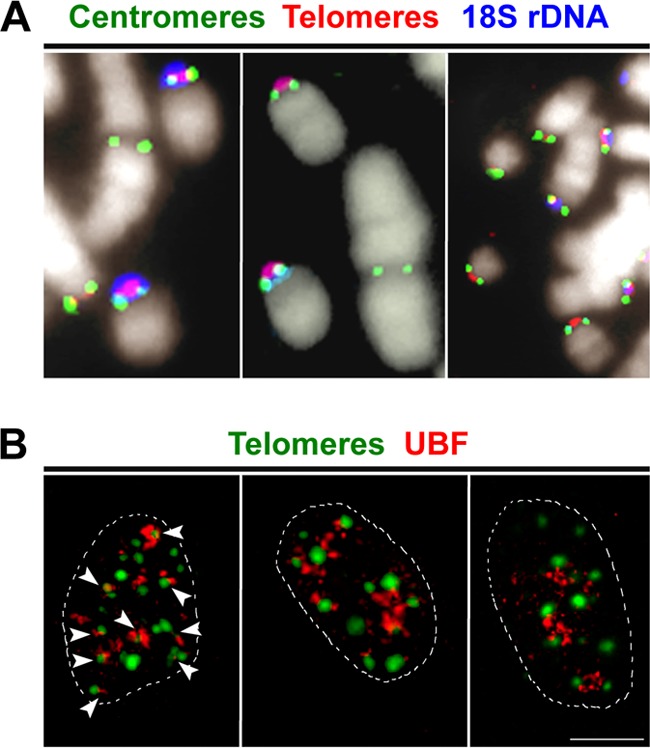
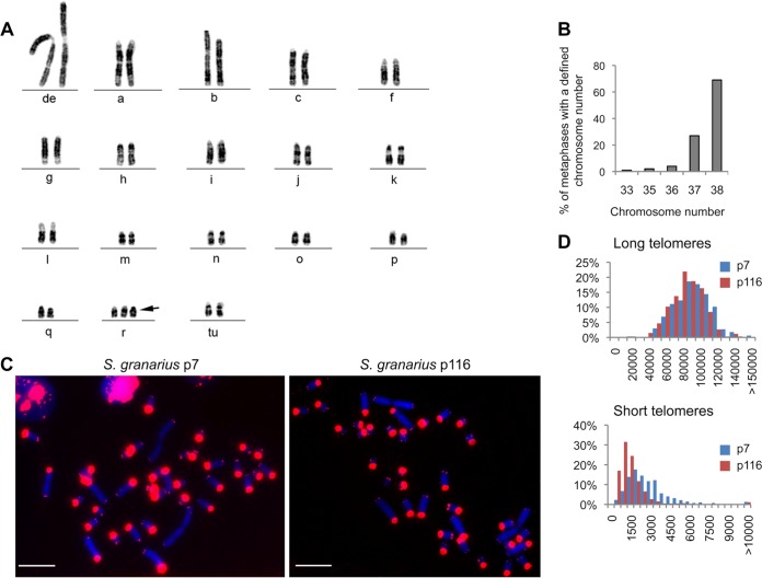
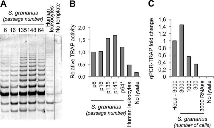
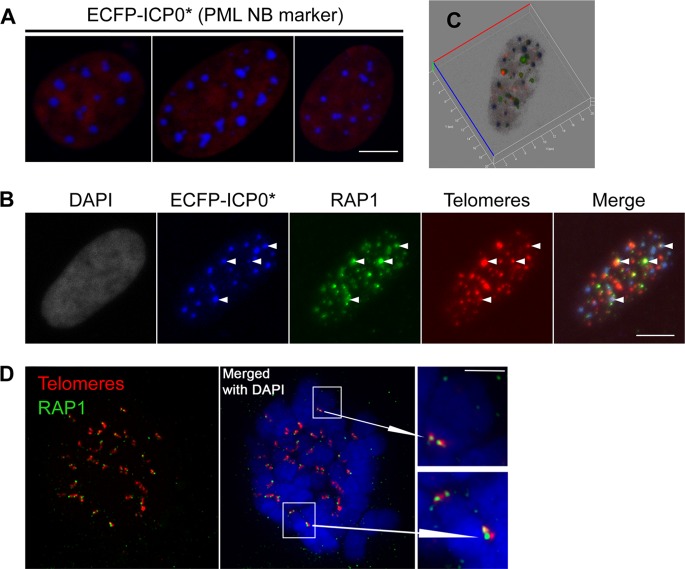
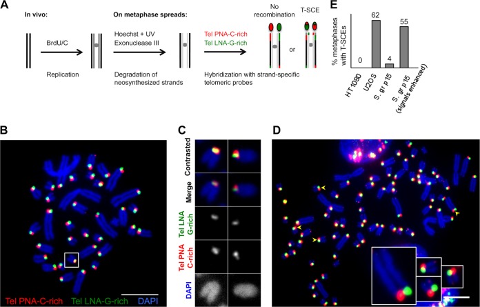
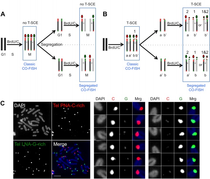
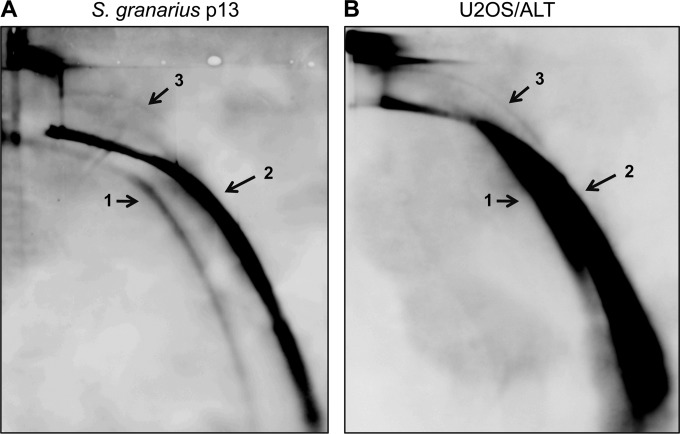
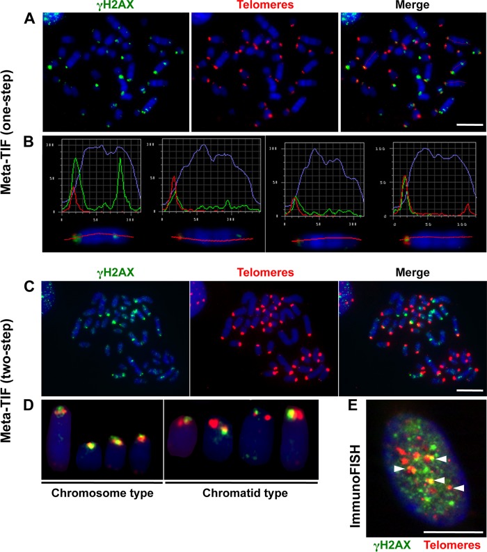
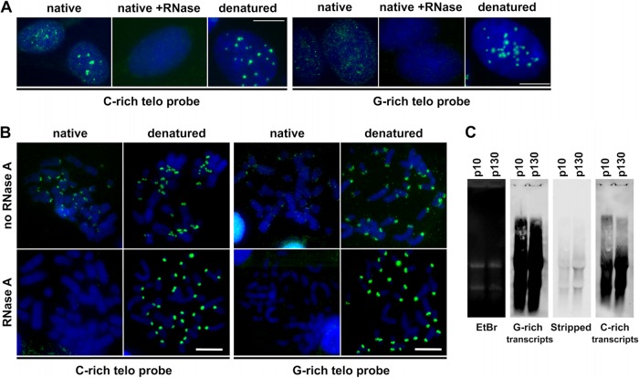
The telomere structure in the Iberian shrew Sorex granarius is characterized by unique, striking features, with short arms of acrocentric chromosomes carrying extremely long telomeres (up to 300 kb) with interspersed ribosomal DNA (rDNA) repeat blocks. In this work, we investigated the telomere physiology of S. granarius fibroblast cells and found that telomere repeats are transcribed on both strands and that there is no telomere-dependent senescence mechanism. Although telomerase activity is detectable throughout cell culture and appears to act on both short and long telomeres, we also discovered that signatures of a recombinogenic activity are omnipresent, including telomere-sister chromatid exchanges, formation of alternative lengthening of telomeres (ALT)-associated PML-like bodies, production of telomere circles, and a high frequency of telomeres carrying marks of a DNA damage response. Our results suggest that recombination participates in the maintenance of the very long telomeres in normal S. granarius fibroblasts. We discuss the possible interplay between the interspersed telomere and rDNA repeats in the stabilization of the very long telomeres in this organism.


Figures

FIG 1
FIG 1
Position of centromeres and nucleolar relationship of acrocentric chromosomes in primary S. granarius fibroblasts. (A) Centromere position on S. granarius acrocentric chromosomes. A two-step experiment was performed, including immunofluorescence assay with ANA-CREST antibodies to detect centromeric proteins (green) and subsequent two-color FISH with a C-rich telomeric PNA probe (Cy3; red) and an 18S rDNA probe (pseudocolored blue). Chromosomes were counterstained with DAPI (pseudocolored white). Centromeres are generally found adjacent to or overlapping signals from telomeric and rDNA probes. (B) Visualization of transcriptionally active nucleoli in primary S. granarius fibroblasts by use of anti-UBF1 antibodies (red). Telomeres were subsequently visualized through PNA FISH (green). In the optical slice at left, arrows indicate close contacts between telomeres and nucleoli. The mean number of nuclear telomere foci (± SEM) was 13.7 ± 2.8 (range, 9 to 26), with 65% ± 2.6% of them being in association with UBF1 signals (n = 90 nuclei). Bar, 10 μm.
FIG 2
FIG 2
Chromosome stability in long-term-culture S. granarius fibroblast cells. (A) Representative karyotype of S. granarius fibroblasts in long-term culture (passage 132; approximately 1 1/2 years). The diploid chromosome complement in female S. granarius is 36, with 32 acrocentric chromosomes and 4 metacentric chromosomes. Arrow, additional chromosome “r.” (B) Chromosome number variation in late-passage fibroblasts (passage 132). Two hundred metaphase spreads were analyzed. (C) Representative images of telomere length analysis by Q-FISH on metaphase chromosomes from primary (passage 7; p7) and long-term (passage 116; p116) fibroblast cultures. The telomeric PNA C-rich probe (Cy3; red) was used, and chromosomes were counterstained with DAPI. The telomere length inequality reported previously for primary fibroblasts (long telomeres on short arms of acrocentric chromosomes versus very short telomeres on all other extremities [10]) was preserved in long-term cultures. Bars, 10 μm. (D) Quantification of Q-FISH telomere intensities for long and short telomeres. See Table 1 for more details.
FIG 3
FIG 3
Fibroblasts of S. granarius contain active telomerase. (A and B) Conventional TRAP analysis of S. granarius fibroblast cells revealed telomerase activity in primary and long-term-culture cells. (A) DNA PAGE gel after conventional TRAP using protein extracts from S. granarius fibroblasts at different passages. A protein extract from PHA-stimulated human leukocytes was used as a positive control. (B) Densitometric analysis of conventional TRAP results. (C) A real-time semiquantitative TRAP assay was performed on cell extracts prepared from early-passage S. granarius cells (passage 9) and compared to the activity found in an equal number of human HeLa cells. Serial dilutions of S. granarius whole-cell extracts (3,000, 1,000, and 300 cell equivalents) resulted in a decreased TRAP activity. Treatment with RNase A and omission of the cell extract served as negative controls.
FIG 4
FIG 4
S. granarius fibroblasts carry APB-like structures. (A) S. granarius primary fibroblasts (passage 7) were transfected with a plasmid expressing an ECFP-ICP0* fusion to reveal putative PML nuclear bodies. The native fluorescence of the ECFP-ICP0* fusion in three transfected nuclei is shown. ICP0* formed 7 to 24 (18 ± 1.94 [mean ± SEM]) large nuclear foci per transfected nucleus (n = 150 nuclei). (B) Cells expressing ECFP-ICP0* were costained with antibodies against the human shelterin protein RAP1 (green) and with a telomeric C-rich PNA probe (Cy3; red) and mounted in Vectashield with DAPI. Maximum-intensity projections are presented for all color channels. The arrows point to close contacts between RAP1, telomeres, and ICP0* (readily visible in the merged image on the far right), suggesting that they are part of the same APB-like structure. (C) 3D reconstitution of the images in panel B. (D) Immuno-FISH on S. granarius metaphase chromosomes, combining antibodies to human RAP1 (green) and a telomeric PNA probe (Cy3; red); chromosomes are stained with DAPI. Bars, 10 μm.
FIG 5
FIG 5
Recombination at telomeres in S. granarius. (A) CO-FISH approach to reveal T-SCEs. After the removal of newly synthesized strands, a C-rich probe will exclusively detect the parental G-rich strand (one-color CO-FISH). If an exchange has taken place after replication, two signals instead of one will be detected at the chromosome extremity. In two-color CO-FISH, strand-specific C-rich and G-rich telomeric probes are used, and T-SCEs are detected as mixed red-green signals. (B) CO-FISH using two strand-specific telomeric probes: TelPNA-C-rich-Cy3 (red) and Tel-LNA-G-rich-FAM (green). Chromosomes were counterstained with DAPI (blue). Only long telomeres present on short acrocentric arms were analyzed. Most extremities show one single green or red robust signal per chromatid. The box indicates a chromosome with mixed signals, indicating a T-SCE. Affected chromosomes varied from metaphase to metaphase. Quantifications of different experiments using one- or two-color CO-FISH are presented in panel E and Table 2. (C) Examples of T-SCEs detected in S. granarius early-passage fibroblasts (p15). (D) Signal enhancement allows the detection of potential highly asymmetric exchanges (very weak green signals colocalizing with the strong red signal, and vice versa [arrowheads]). Enlarged examples are presented. The segregated CO-FISH analysis presented in Fig. 6 indicates that such colocalizations correspond to bona fide T-SCEs. Bars, 10 μm. (E) Quantification of T-SCEs in S. granarius early-passage (S. gr p15) fibroblasts relative to U2OS/ALT and HT1080/TEL+ human cancer cell lines (n = 30 metaphase spreads for each condition). The frequency of metaphase chromosomes carrying T-SCEs when only “robust” fluorescence signals are taken into account appears to be low in S. granarius fibroblasts. However, when T-SCEs are searched upon enhancement of signals, the frequency is much higher. The fact that these are bone fide T-SCEs was confirmed by segregated CO-FISH analysis (Fig. 6).
FIG 6
FIG 6
(A) BrdU/C incorporation during two cell cycles prior to the CO-FISH procedure, using strand-specific telomeric probes, results in segregation of the unsubstituted G- and C-rich strands into different chromosomes, such that during the second M phase, every chromosome extremity will be stained, after the CO-FISH procedure, with only one probe, either red (TelPNA-C-rich-Cy3) or green (TelLNA-G-rich-FAM). (B) If a T-SCE occurs during the first cell cycle, the two unsubstituted strands will cosegregate during the first mitosis and will be detected on different sister chromatids of the same chromosome during the second M phase. The CO-FISH procedure will then reveal one red and one green signal on the same chromosome extremity (marked 1). If a T-SCE occurs during the second cell cycle, this exchange will result in same-color doublets, either red or green (marked 2). However, if a second exchange affects an extremity that had already undergone T-SCE during the first cell cycle, doublets will be of both colors (marked 1&2). (C) Two-color segregated CO-FISH in S. granarius. Highly asymmetric two-color doublets are frequently detected in S. granarius fibroblasts (p15). Examples of such T-SCEs are enlarged and color decomposed on the right. The TelPNA-C-rich-Cy3 probe is more efficient than the TelLNA-G-rich-FAM probe for detecting small doublets. Chromosomes were counterstained with DAPI (blue). Bar, 10 μm.
FIG 7
FIG 7
Telomeric circles are detected in primary S. granarius fibroblasts. Ten micrograms of genomic DNA from S. granarius early-passage (p13) fibroblasts (A) and 20 μg genomic DNA from the human ALT cancer cell line U2OS (B) were digested with MboI, separated by 2D gel electrophoresis, transferred onto N+ nylon membranes, and hybridized with a digoxigenin-labeled telomeric C-rich oligonucleotide. Arrow 1, single-stranded linear DNA; arrow 2, double-stranded linear DNA; arrow 3, circular DNA.
FIG 8
FIG 8
Spontaneous telomere dysfunction in primary S. granarius fibroblasts. (A) Meta-TIF analysis of S. granarius fibroblasts (one-step protocol). Metaphase chromosomes were first stained with anti-γH2AX antibodies (green) and subsequently stained with a telomeric PNA probe (red) for detection of telomere-induced foci (TIF). Chromosomes were counterstained with DAPI (blue). (B) Relative green and red fluorescence intensities of particular chromosomes from the metaphase spread shown in panel A, illustrating either perfect colocalization of red and green signals or the spread of green signals toward the interstitial region. (C) Meta-TIF analysis of S. granarius fibroblasts by a two-step protocol involving immunofluorescence assay with anti-γH2AX antibodies, with image acquisition (green; left panel), as well as hybridization with a telomeric PNA probe, with visualization (red; middle panel). The right panel show the merge of the two visualization steps. (D) Illustration of chromosome- and chromatid-type TIF detected in a two-step experiment. The images show staining with anti-γH2AX antibodies (green) and a telomeric PNA probe (red). A quantification of these experiments is presented in Table 3. (E) Detection of TIF in S. granarius interphase nucleus by a one-step protocol. The image shows staining with anti-γH2AX antibodies (green) and a telomeric PNA probe (red). Bars, 10 μm.
FIG 9
FIG 9
Both G-rich and C-rich telomere strands are expressed in S. granarius cells. (A) RNA FISH experiments with strand-specific probes on S. granarius interphase nuclei. The C-rich probe detected either the UUAGGG or TTAGGG sequence, and the G-rich probe detected either CCCUAA or CCCTAA, depending on whether the hybridization was done on native or denatured nuclei, respectively. Signals were visible with both probes under native conditions without RNase treatment, albeit at lower intensity when the probe was G-rich. The signal was completely lost when native preparations were treated with RNase. Both probes detected telomeres equally efficiently under denaturing conditions. Nuclei are stained with DAPI (blue). (B) Similar RNA FISH experiment on metaphase chromosome preparations obtained by cytospin centrifugation under native or denatured conditions and treated or not treated with RNase. Once again, the C-rich probe yielded stronger signals than the G-rich probe, only on native, RNase-untreated chromosomes. Bars, 10 μm. (C) Northern blot analysis of S. granarius telomeric RNA transcripts in cells at different passages (p). (Left) Visualization of total RNA in the gel by ethidium bromide (EtBr) staining before transfer onto a membrane. (Second panel) Hybridization with a C-rich telomere probe labeled with digoxigenin. (Third panel) Verification of the stripping efficiency. (Right) Hybridization with a G-rich probe labeled with digoxigenin.

References

    1. Pfeiffer V, Lingner J. 2013. Replication of telomeres and the regulation of telomerase. Cold Spring Harb. Perspect. Biol. 5:a010405. 10.1101/cshperspect.a010405 - DOI - PMC - PubMed
    1. Tumpel S, Rudolph KL. 2012. The role of telomere shortening in somatic stem cells and tissue aging: lessons from telomerase model systems. Ann. N. Y. Acad. Sci. 1266:28–39. 10.1111/j.1749-6632.2012.06547.x - DOI - PubMed
    1. Hug N, Lingner J. 2006. Telomere length homeostasis. Chromosoma 115:413–425. 10.1007/s00412-006-0067-3 - DOI - PubMed
    1. Pickett HA, Cesare AJ, Johnston RL, Neumann AA, Reddel RR. 2009. Control of telomere length by a trimming mechanism that involves generation of t-circles. EMBO J. 28:799–809. 10.1038/emboj.2009.42 - DOI - PMC - PubMed
    1. Griffith JD, Comeau L, Rosenfield S, Stansel RM, Bianchi A, Moss H, de Lange T. 1999. Mammalian telomeres end in a large duplex loop. Cell 97:503–514. 10.1016/S0092-8674(00)80760-6 - DOI - PubMed

Publication types

LinkOut - more resources