. 2014 Jun 3;111(22):8161-6.
doi: 10.1073/pnas.1318355111. Epub 2014 May 19.

Ultrasound-guided tissue fractionation by high intensity focused ultrasound in an in vivo porcine liver model

Affiliations

Ultrasound-guided tissue fractionation by high intensity focused ultrasound in an in vivo porcine liver model

Tatiana D Khokhlova et al. Proc Natl Acad Sci U S A. .

Abstract

The clinical use of high intensity focused ultrasound (HIFU) therapy for noninvasive tissue ablation has been recently gaining momentum. In HIFU, ultrasound energy from an extracorporeal source is focused within the body to ablate tissue at the focus while leaving the surrounding organs and tissues unaffected. Most HIFU therapies are designed to use heating effects resulting from the absorption of ultrasound by tissue to create a thermally coagulated treatment volume. Although this approach is often successful, it has its limitations, such as the heat sink effect caused by the presence of a large blood vessel near the treatment area or heating of the ribs in the transcostal applications. HIFU-induced bubbles provide an alternative means to destroy the target tissue by mechanical disruption or, at its extreme, local fractionation of tissue within the focal region. Here, we demonstrate the feasibility of a recently developed approach to HIFU-induced ultrasound-guided tissue fractionation in an in vivo pig model. In this approach, termed boiling histotripsy, a millimeter-sized boiling bubble is generated by ultrasound and further interacts with the ultrasound field to fractionate porcine liver tissue into subcellular debris without inducing further thermal effects. Tissue selectivity, demonstrated by boiling histotripsy, allows for the treatment of tissue immediately adjacent to major blood vessels and other connective tissue structures. Furthermore, boiling histotripsy would benefit the clinical applications, in which it is important to accelerate resorption or passage of the ablated tissue volume, diminish pressure on the surrounding organs that causes discomfort, or insert openings between tissues.


Conflict of interest statement

The authors declare no conflict of interest.

Figures

Fig. 1.
Fig. 1.
Illustration of the experimental setup and the physical principle of the HIFU-induced boiling-histotripsy procedure. (A) A hand-held 2 MHz HIFU transducer (Inset), was attached to a water-filled coupling cone, that was brought into contact with exposed liver of a 100- to 150-lb domestic swine. (Scale bar: 1 cm.) The ultrasound imaging probe was inserted through the central opening of the transducer for targeting and monitoring of the exposure. The HIFU focus was placed at depths of 5–18 mm below the tissue surface. (B) The idea behind boiling-histotripsy exposures is to make use of highly nonlinear ultrasound waves that form at the HIFU focus and contain shock fronts. Ultrasound absorption at the shock fronts in tissue is very efficient, which leads to temperature elevations up to 100 °C in a matter of milliseconds in a very small volume around the focus, where shocks are present. A large boiling bubble then forms at the HIFU focus, and its interaction with the remainder of the incident HIFU pulse leads to tissue fractionation. To avoid thermal effects on tissue, short HIFU pulses (1–10 ms) and long intervals between the pulses (0.1–1 s) were used. Each exposure lasted for 50 s, and then the transducer was moved to another spot on the liver. (C) Explosive growth of the millimeter-sized boiling bubble and its interaction with the HIFU field causes the breakdown of tissue into submicrometer fragments. The hypothesized physical mechanism behind tissue fractionation is the formation of a miniature acoustic fountain within the boiling bubble and ultrasound-induced tissue atomization.
Fig. 2.
Fig. 2.
B-mode ultrasound images taken throughout a boiling-histotripsy exposure. The hypoechoic line at the top of the images corresponds to the interface between the tissue and water-filled coupling cone of the HIFU transducer. The first frame illustrates the position of the HIFU focus inside the liver. The pulsing protocol in this particular case was as follows: HIFU pulse duration 10 ms, pulse repetition frequency 1 s, and treatment duration 50 s. After the first HIFU pulse was delivered, a bright hyperechoic region (indicated by the white arrow) appeared at the HIFU focus. Its brightness was the highest immediately after the pulse was delivered, and decreased slightly before the next pulse arrived. The hyperechoic region continued to grow within the next 20 s of exposure, and then its size saturated. During the subsequent 30 s, the size of the hyperechoic region did not change, which is consistent with our previous observations in ex vivo tissues. After the last HIFU pulse was delivered, the hyperechoic region persisted for several seconds (last frame), but eventually faded completely, and the treated region was indistinguishable from the surrounding tissue. However, the maximum size of the hyperechoic region corresponds well (within the resolution of the ultrasound imaging system) to the size of the resulting lesion (Figs. 3 and 4).
Fig. 3.
Fig. 3.
Representative gross photographs of the boiling-histotripsy lesions induced in ex vivo (A, B, E, and F) and in vivo (C and D) porcine liver by using different HIFU pulsing protocols, in which the duty factor was kept constant (0.01) and the HIFU pulse duration varied (1–500 ms). (A) When short (less than 20 ms) HIFU pulses were used in the ex vivo setting, the outcome was a void filled with liquid of the same color as surrounding tissue, with no signs of thermal denaturation. The liquid could be easily removed (B), and the walls of the remaining cavity repeated the shape of liver lobules (indicated by yellow arrows). The same HIFU treatment delivered in vivo (C) created a void of about the same size as ex vivo, but the liquefied contents were much darker and redder than the surrounding tissue. With the contents removed, the lesion structure also repeated the structure of the liver lobules (yellow arrows). (D) As the HIFU pulse length increased (20–100 ms) in the ex vivo setting, the lesion contents turned into a thick white paste and the lesion edges were blanched (E). In vivo, the thermal damage was not as obvious; the lesion looked essentially the same as the one shown in C. With a further increase in pulse duration (100–500 ms), both in vivo and ex vivo, the lesion turned into a well-defined area of coagulative necrosis containing large vacuoles (F).
Fig. 4.
Fig. 4.
Representative histological sections of the frozen samples of boiling-histotripsy lesions, produced in in vivo (Upper) or ex vivo (Lower) porcine liver, stained for NADH-d. The NADH-d stain shows the presence (stained purple) or absence (unstained) of enzymatic activity in tissue, which is used as an indication of thermal damage. Stromal tissue also remains unstained, which allows for visualization of the lobular structure of liver tissue. One of the lobules in A is outlined by a red dotted line, and all of the lesions are outlined by dotted yellow lines. (A) Liquid lesions produced by using 10-ms-long HIFU pulses. Ice crystal formation in the lesions, both in vivo and ex vivo, indicates that the lesion contents are liquefied. There is no indication of thermal damage, neither to the lesion contents, nor to the surrounding tissue. The higher magnification images stained with H&E (Fig. S6A) indicate that the tissue debris within the lesions are no larger than the cell nucleus. The sizes of the in vivo and ex vivo lesions are similar, and the shapes depend on the lobular structure of tissue: The lesions are “contained” by connective tissue, in accord with gross observations. (B) “Paste” lesions produced by using longer (100 ms) HIFU pulses. The contents of both lesions are liquefied, but thermal denaturation of the lesion contents occurs to a greater extent in vivo than ex vivo. In vivo, the lesion contents are completely thermally denatured, as are the lesion boundaries within a 100-μm margin. Ex vivo, the thermal effects are confined to the small areas adjacent to the connective tissue (blue arrows). The in vivo lesion also contains a continuous area of thermal necrosis with large vacuoles, outlined with blue dotted line. The size of the intact tissue fragments, contained in both lesions, is larger (up to 50 μm) than in the liquid lesion (Fig. 6B). (C) Vacuolated thermal lesions produced by using a single 500-ms-long HIFU pulse. The size, structure, and degree of thermal damage are very similar in the in vivo and ex vivo lesions. Both lesions contain thermally necrosed tissue with large vacuoles left by the boiling bubbles. The tissue next to the vacuoles is squeezed and disrupted, whereas closer to the lesion border, it appears thermally fixed. Surprisingly, some of the lesions contain viable cells within the blood vessels, as indicated by the black arrows. (Scale bars: 500 μm.)
Fig. 5.
Fig. 5.
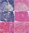
Higher magnification histological images stained with NADH-d and H&E demonstrating the important details of the liquid lesions produced in vivo by boiling histotripsy. (A) H&E (Left) and NADH-d (Right) stained histological images of a liquid boiling histotripsy lesion. Connective tissue, located within the liquid boiling histotripsy lesions, frequently gets thermally damaged (dotted yellow line), whereas the hepatocytes and the debris thereof are not thermally denatured. If, however, a connective tissue structure (e.g., a blood vessel or a biliary system element) is located immediately adjacent to the lesion, it is not affected (blue arrows) either mechanically or thermally. (B) Multifocal boiling-histotripsy lesion produced under the conditions of significant tissue movement due to breathing and heartbeat. The HIFU pulsing protocol used was designed to produce a single liquid lesion (pulse duration 10 ms, pulse repetition frequency 1 Hz, 50-s duration), but because of tissue motion, the HIFU focus location changed from pulse to pulse, and small liquefied areas formed at several places, but did not merge. Tissue motion is a challenge in boiling histotripsy, which can be addressed by gating the HIFU pulses by breathing or cardiac motion. (C) Lysed RBCs are abundant in the liquid boiling-histotripsy lesions and are seen in the H&E-stained sections of the lesions as a red tint (left side of the image). Intact RBCs were not observed within the lesions, indicating that the blood inflow to the emulsified void continued only through the HIFU exposure and stopped shortly afterward. (D) High magnification H&E stained image of the liquid lesion border, which is very sharp and only one to two cells (10–20 μm) in width. (Scale bars: A and B, 500 μm; C, 100 μm.)
Fig. 6.
Fig. 6.
Magnified view of the H&E-stained histological sections of the debris contained in liquid (A) and paste (B) lesions produced in vivo. Ice crystal formation is pronounced in both images, indicating that the lesion contents is mostly liquefied. Both lesions contain a few intact cell nuclei (blue arrows), that are much more abundant in the paste lesion. The latter also contains larger tissue fragments, up to 100 μm in size, outlined by yellow dotted lines. (Scale bars: 50 μm.)

Similar articles

Cited by

References

    1. Dubinsky TJ, Cuevas C, Dighe MK, Kolokythas O, Hwang JH. High-intensity focused ultrasound: Current potential and oncologic applications. AJR Am J Roentgenol. 2008;190(1):191–199. - PubMed
    1. Bailey MR, Khokhlova VA, Sapozhnikov OA, Kargl SG, Crum LA. Physical mechanisms of the therapeutic effect of ultrasound (a review) Acoust Phys. 2003;49:369–388.
    1. Leslie T, et al. High-intensity focused ultrasound treatment of liver tumours: Post-treatment MRI correlates well with intra-operative estimates of treatment volume. Br J Radiol. 2012;85(1018):1363–1370. - PMC - PubMed
    1. Crouzet S, et al. Whole-gland ablation of localized prostate cancer with high-intensity focused ultrasound: Oncologic outcomes and morbidity in 1002 patients. Eur Urol. 2014;65(5):907–914. - PubMed
    1. Ren XL, et al. Extracorporeal ablation of uterine fibroids with high-intensity focused ultrasound: Imaging and histopathologic evaluation. J Ultrasound Med. 2007;26(2):201–212. - PubMed

Publication types

MeSH terms