. 2014 Jun 3;111(22):8025-30.
doi: 10.1073/pnas.1401180111. Epub 2014 May 19.

Structure of the pseudokinase-kinase domains from protein kinase TYK2 reveals a mechanism for Janus kinase (JAK) autoinhibition

Affiliations

Structure of the pseudokinase-kinase domains from protein kinase TYK2 reveals a mechanism for Janus kinase (JAK) autoinhibition

Patrick J Lupardus et al. Proc Natl Acad Sci U S A. .

Abstract

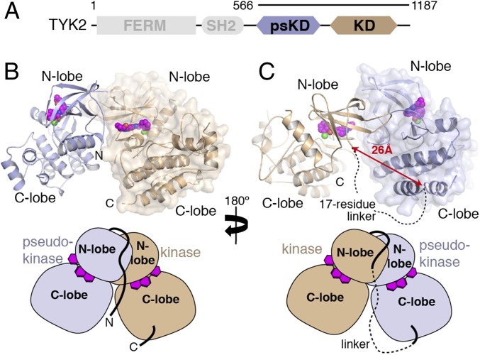
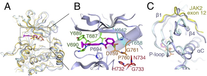
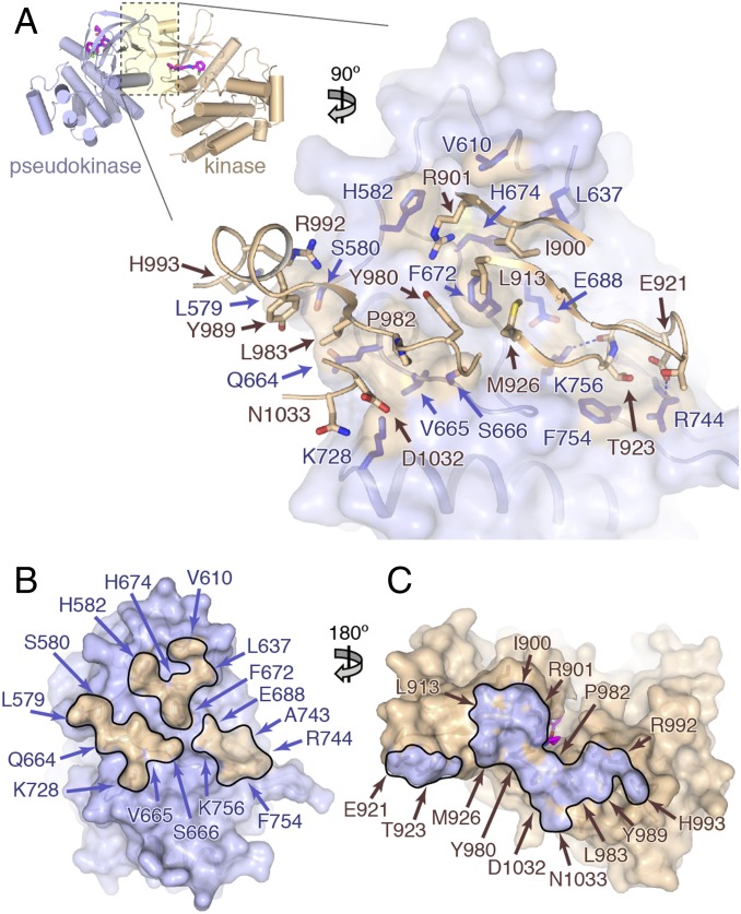
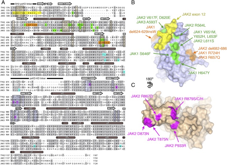
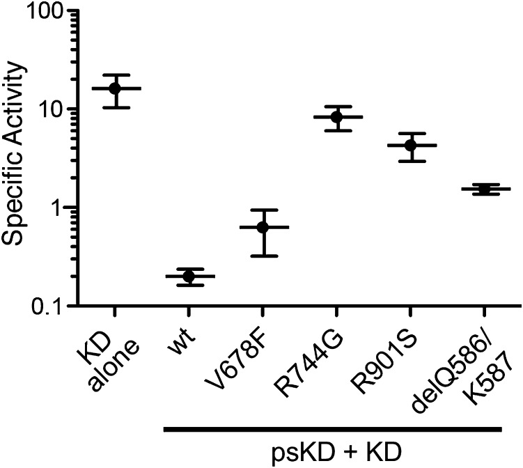
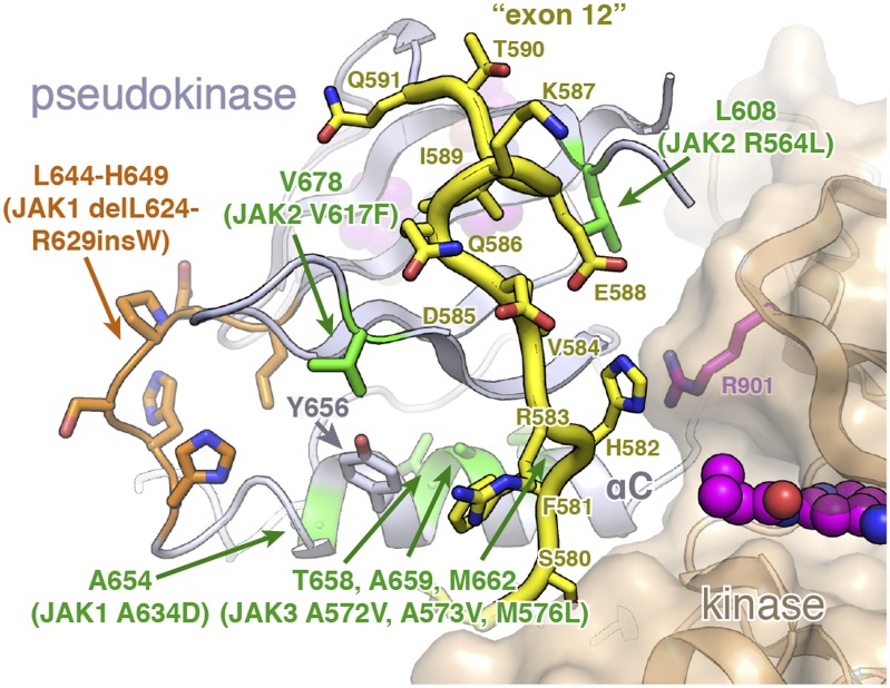
Janus kinases (JAKs) are receptor-associated multidomain tyrosine kinases that act downstream of many cytokines and interferons. JAK kinase activity is regulated by the adjacent pseudokinase domain via an unknown mechanism. Here, we report the 2.8-Å structure of the two-domain pseudokinase-kinase module from the JAK family member TYK2 in its autoinhibited form. We find that the pseudokinase and kinase interact near the kinase active site and that most reported mutations in cancer-associated JAK alleles cluster in or near this interface. Mutation of residues near the TYK2 interface that are analogous to those in cancer-associated JAK alleles, including the V617F and "exon 12" JAK2 mutations, results in increased kinase activity in vitro. These data indicate that JAK pseudokinases are autoinhibitory domains that hold the kinase domain inactive until receptor dimerization stimulates transition to an active state.

Keywords: JAK1; JAK3.


Conflict of interest statement

The authors declare no conflict of interest.

Figures

Fig. 1.
Fig. 1.
Crystal structure of the TYK2 pseudokinase–kinase. (A) Simplified domain structure of TYK2 indicating the boundaries of the pseudokinase–kinase construct used in this study. (B) Diagram of the pseudokinase (blue) and kinase (tan) complex. From this view, the N and C termini of the crystallized construct are visible. Protein backbone is shown with the all-atom surface of the kinase domain displayed to aid consideration of the interaction interface. The kinase inhibitor molecule compound 7012 is shown in the active site as spheres (carbon atoms colored purple). (C) The diagram from B is rotated 180°, with pseudokinase and kinase represented in the same color, but the surface of the pseudokinase is shown. In this view, the region of the interdomain linker between the pseudokinase and kinase are visible, with a distance measurement and a dashed line shown to illustrate the connection made by the unobserved peptide linkage. Simplified diagrams are shown beneath each panel for orientation.
Fig. 2.
Fig. 2.
Comparison of the TYK2, JAK1, and JAK2 pseudokinases. (A) Overlay of the TYK2 pseudokinase domain (blue) with the pseudokinase from JAK1 (PDB ID code 4L00; gold) and JAK2 (PDB ID code 4FVQ; cyan). Protein chain is shown as a cartoon model, with ligands shown as sticks (TYK2-bound compound 7012 in magenta, JAK2-bound ATP in yellow). (B) Close-up view of the TYK2 pseudokinase active site. Compound 7012 is shown in magenta, and the positions of canonical kinase features colored [hinge: green; HGN (∼“HRD”) motif: red; DPG (∼“DFG”) motif: orange; αC-helix salt bridge residues: cyan]. (C) View of the exon 12 N-terminal segment of JAK2 compared with TYK2. Protein chains are colored as in A, with the JAK2 exon 12 segment colored yellow.
Fig. 3.
Fig. 3.
The TYK2 pseudokinase–kinase interaction. (A) (Upper) Overview of the pseudokinase–kinase complex. The pseudokinase is colored in blue, and kinase is colored in tan. (Lower) Zoomed-in view of the pseudokinase–kinase interface. The pseudokinase is shown facing up as a blue semitransparent molecular surface, with the kinase in front shown as tan worm and stick side chains. Pseudokinase residues within 4.5 Å of the kinase domain have side chains displayed as dark blue sticks (with tan surface). (B and C) Open-book view of the pseudokinase–kinase interface. Surface representations of the pseudokinase (B) and kinase (C) domains, colored as in A with surfaces within 4.5 Å of the partner domain outlined and highlighted the opposite color. Residues found in the interface are labeled.
Fig. 4.
Fig. 4.
Cancer-associated JAK alleles map to the TYK2 pseudokinase–kinase interface. (A) Sequence alignment of the four human JAKs, with cancer-associated JAK alleles colored according to the type of mutation. For the pseudokinase, point mutants are shown in green, deletions are shown in orange, and the exon 12 segment is shown in yellow. For the kinase, point mutants are shown in magenta. Mutations outside the N-lobes of the pseudokinase or kinase are shown in cyan. Residues found within the pseudokinase and kinase interface are boxed and shaded in tan and blue, respectively. Protein secondary structure is also shown above the sequences, with α-helices shown as cylinders and β-sheets shown as rectangular arrows. (B and C) Open-book surface views of the pseudokinase (B) and kinase (C) domains, with select JAK alleles mapped onto the models. Mutations are colored as in A. The perspective shown is identical to that displayed in Fig. 3 B and C.
Fig. 5.
Fig. 5.
TYK2 pseudokinase–kinase mutants analogous to cancer-associated JAK alleles are active in vitro. The activity of the kinase domain of wild-type TYK2 (KD, residues 885–1176) was compared with the pseudokinase and kinase domains of wild-type TYK2 (wt, residues 566–1187) as well as a number of pseudokinase–kinase interface mutants (V678F, R744G, R901S, and delQ586/K587). Specific activity was measured in an assay monitoring phosphorylation of a synthetic peptide derived from the JAK3 sequence and calculated based on the percentage conversion to phosphorylated product over time and the concentration of TYK2 used. Values shown have units of nanomolar concentration of product formed per minute per nanomolar concentration of TYK2 and are the mean of more than five measurements ± SD.
Fig. 6.
Fig. 6.
V617 and αC-helix alleles carry “second-shell” mutations that may disrupt the exon 12 segment. A close-up view of the TYK2 N terminus, which corresponds to the JAK2 exon 12 segment that is frequently mutated in MPN disorders. The TYK2 N-terminal segment is highlighted in yellow with side chains shown as sticks. Point mutations in this region are highlighted in green, with a JAK1 deletion highlighted in orange.

References

    1. Leonard WJ, O’Shea JJ. Jaks and STATs: Biological implications. Annu Rev Immunol. 1998;16:293–322. - PubMed
    1. Haan C, Kreis S, Margue C, Behrmann I. Jaks and cytokine receptors—an intimate relationship. Biochem Pharmacol. 2006;72(11):1538–1546. - PubMed
    1. Rodig SJ, et al. Disruption of the Jak1 gene demonstrates obligatory and nonredundant roles of the Jaks in cytokine-induced biologic responses. Cell. 1998;93(3):373–383. - PubMed
    1. Parganas E, et al. Jak2 is essential for signaling through a variety of cytokine receptors. Cell. 1998;93(3):385–395. - PubMed
    1. Neubauer H, et al. Jak2 deficiency defines an essential developmental checkpoint in definitive hematopoiesis. Cell. 1998;93(3):397–409. - PubMed

Publication types

MeSH terms

Associated data