. 2014 Jun 3;111(22):7968-73.
doi: 10.1073/pnas.1310842111. Epub 2014 May 19.

Fluid shear stress threshold regulates angiogenic sprouting

Affiliations

Fluid shear stress threshold regulates angiogenic sprouting

Peter A Galie et al. Proc Natl Acad Sci U S A. .

Abstract

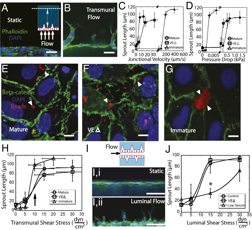
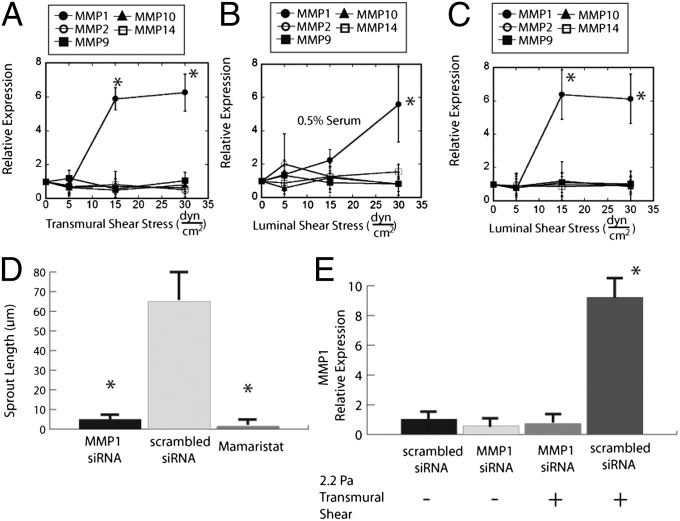
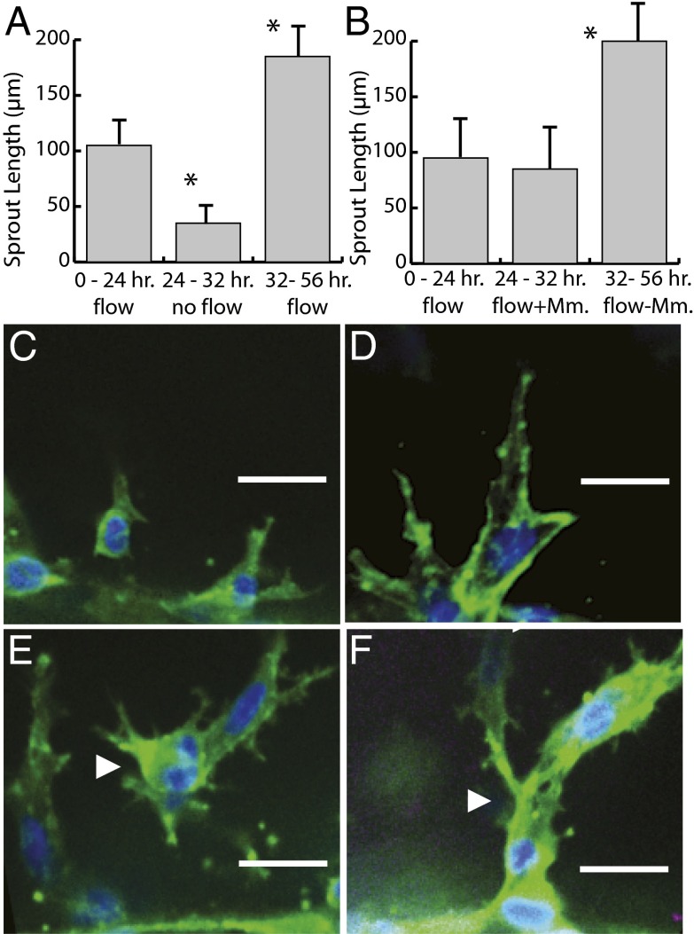
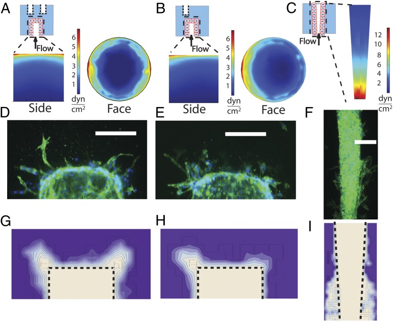
The density and architecture of capillary beds that form within a tissue depend on many factors, including local metabolic demand and blood flow. Here, using microfluidic control of local fluid mechanics, we show the existence of a previously unappreciated flow-induced shear stress threshold that triggers angiogenic sprouting. Both intraluminal shear stress over the endothelium and transmural flow through the endothelium above 10 dyn/cm(2) triggered endothelial cells to sprout and invade into the underlying matrix, and this threshold is not impacted by the maturation of cell-cell junctions or pressure gradient across the monolayer. Antagonizing VE-cadherin widened cell-cell junctions and reduced the applied shear stress for a given transmural flow rate, but did not affect the shear threshold for sprouting. Furthermore, both transmural and luminal flow induced expression of matrix metalloproteinase 1, and this up-regulation was required for the flow-induced sprouting. Once sprouting was initiated, continuous flow was needed to both sustain sprouting and prevent retraction. To explore the potential ramifications of a shear threshold on the spatial patterning of new sprouts, we used finite-element modeling to predict fluid shear in a variety of geometric settings and then experimentally demonstrated that transmural flow guided preferential sprouting toward paths of draining interstitial fluid flow as might occur to connect capillary beds to venules or lymphatics. In addition, we show that luminal shear increases in local narrowings of vessels to trigger sprouting, perhaps ultimately to normalize shear stress across the vasculature. Together, these studies highlight the role of shear stress in controlling angiogenic sprouting and offer a potential homeostatic mechanism for regulating vascular density.

Keywords: angiogenesis; force; mechanotransduction; migration; morphogenesis.


Conflict of interest statement

The authors declare no conflict of interest.

Figures

Fig. 1.
Fig. 1.
(A and B) Schematic and representative image of a mature endothelial cell monolayer plated on the surface of a collagen gel (A) and sprouting in response to perfusion with transmural flow (B). (Scale bar, 50 µm.) (C and D) Mean sprout length of mature untreated and VEΔ monolayers and immature monolayers exposed to transmural flow of several junctional velocities (C) and pressure drops (D). Linear ANOVAs followed by Tukey tests were used to determine that the sprouting responses of control and VEΔ groups were significantly different (*P < 0.05). (E–G) Monolayer of mature untreated (E) and VEΔ (F) and immature (G) conditions. Green, β-catenin; blue, nucleus; red, 1-µm beads. The white arrowheads point to the distributed beads accumulated on the monolayer surface. (Scale bar, 10 µm.) (H) Mean sprout length as a function of transmural shear for nontreated mature, VEΔ mature, and immature monolayers. Again, post hoc Tukey tests were conducted to calculate the significant difference in the reduced serum group (*P < 0.05). (I) Surface of cylindrical channel for static (I, i) and luminal flow (I, ii) conditions. Green, F-actin; blue, nucleus. (Scale bar, 100 µm.) (J) Mean sprout length as a function of luminal shear for nontreated and VEΔ cells and cells exposed to 0.5% serum.
Fig. 2.
Fig. 2.
(A) MMP message levels for MMP1, MMP2, MMP9, MMP10, and MMP14 double normalized to GAPDH and static controls in response to transmural shear stress. Two-sample t tests were performed for each MMP primer to determine statistical significance between the applied shear and static controls (*P < 0.05). (B) MMP message levels for cells exposed to luminal flow and reduced serum (*P < 0.05). (C) MMP message levels in response to luminal flow (*P < 0.05). (D) Mean sprout length for cells treated with MMP1 DsiRNA, a scrambled DsiRNA control, and Mamaristat. Two-sample t tests were calculated to test for a significant change from untreated controls (*P < 0.05). (E) Validation of successful knockdown by qRT-PCR. Transmural flow only significantly increases message levels of MMP1 (*P < 0.05).
Fig. 3.
Fig. 3.
(A and B) Mean sprout length after stopping and restarting fluid flow (A) and temporary Mamaristat treatment (B). Two-sample t tests were calculated to determine a significant change from the initial 24-h sprouting response (*P < 0.05). (C and D) Representative image of sprouts after 8 h of static conditions (C) or Mamaristat treatment (D). Green, F-actin; blue, nucleus. (Scale bar, 50 µm.) (E and F) Representative image of sprouts after the 24-h period following resumption of flow (E) or washing out of Mamaristat (F). White arrowheads point to one of the single-cell sprouts observed in the halted-flow group (E) and to the multicellular structures observed in the continuous-flow group (F). Green, F-actin; blue, nucleus. (Scale bar, 50 µm.)
Fig. 4.
Fig. 4.
(A–C) Finite-element model predictions of shear stress for the two-void, transmural flow configuration (A), the single-void configuration (B), and the endothelial-lined nozzle (C). (D–F) Representative images of the flow-induced sprouting for the three configurations: two-void (D), single-void (E), and nozzle (F). Green, F-actin; blue, nucleus. (Scale bar, 200 µm.) (G–I) Frequency plots generated by at least five images of sprouting in the three configurations: two-void (G), single-void (H), and nozzle (I).

References

    1. Tuder RM, Flook BE, Voelkel NF. Increased gene expression for VEGF and the VEGF receptors KDR/Flk and Flt in lungs exposed to acute or to chronic hypoxia. Modulation of gene expression by nitric oxide. J Clin Invest. 1995;95(4):1798–1807. - PMC - PubMed
    1. Potente M, Gerhardt H, Carmeliet P. Basic and therapeutic aspects of angiogenesis. Cell. 2011;146(6):873–887. - PubMed
    1. Kuo L, Davis MJ, Chilian WM. Endothelium-dependent, flow-induced dilation of isolated coronary arterioles. Am J Physiol. 1990;259(4 Pt 2):H1063–H1070. - PubMed
    1. Dawson JM, Hudlická O. The effects of long term administration of prazosin on the microcirculation in skeletal muscles. Cardiovasc Res. 1989;23(11):913–920. - PubMed
    1. Egginton S, Zhou AL, Brown MD, Hudlická O. Unorthodox angiogenesis in skeletal muscle. Cardiovasc Res. 2001;49(3):634–646. - PubMed

Publication types

MeSH terms

Substances