

Nd:YAG membranotomy for preretinal hemorrhage secondary to valsalva retinopathy
- PMID: 24843309
- PMCID: PMC4023117
- DOI: 10.1016/j.sjopt.2014.02.006
Nd:YAG membranotomy for preretinal hemorrhage secondary to valsalva retinopathy
Abstract
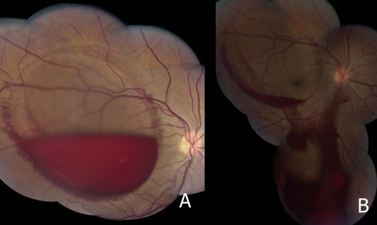
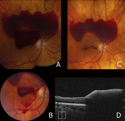
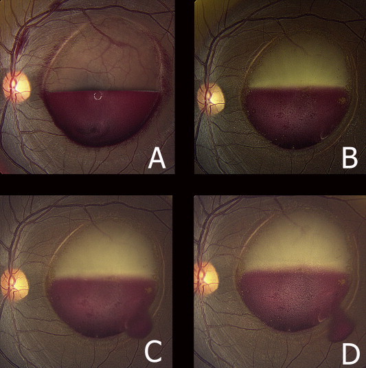
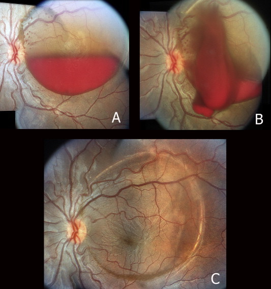
Purpose: To report four cases of premacular hemorrhage secondary to valsalva retinopathy treated with Nd:YAG membranotomy and discuss techniques as well as the literature.
Design: Retrospective case series.
Methods: A retrospective review was conducted for four patients with vision obstructing hemorrhage secondary to valsalva retinopathy. These patients were all treated with Nd:YAG membranotomy.
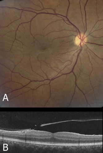
Results: Four patients with premacular hemorrhage secondary to valsalva retinopathy were treated with Nd:YAG laser creating a membranotomy to drain the hemorrhage. Power settings ranged from 1.7 to 3.8 mJ. Visual acuity at presentation ranged from 20/400 (1 patient) to count fingers (3 patients). Visual acuity improved in three out of four patients after laser treatment. Final visual acuity ranged from 20/20 to 20/30 in these three patients. One patient was lost to follow up after performing laser membranotomy and therefore visual acuity after treatment was not obtained. No complications were noted.
Conclusion: Nd:YAG membranotomy is a non-invasive, office-based treatment option that may be successfully used to treat premacular hemorrhage secondary to valsalva retinopathy.
Keywords: Premacular hemorrhage; Valsalva retinopathy; YAG membranotomy.
Figures

References
-
- Celebi S., Kukner A.S. Photodisruptive Nd:YAG laser in the management of premacular subhyaloid hemorrhage. Eur J Ophthalmol. 2001;11(3):281–286. - PubMed
-
- Khan M.T., Saeed M.U., Shehzad M.S., Qazi Z.A. Nd:YAG laser treatment for valsalva premacular hemorrhages: 6 month follow up: alternative management options for preretinal premacular hemorrhages in Valsalva retinopathy. Int Ophthalmol. 2008;28(5):325–327. - PubMed
-
- Kwok A.K., Lai T.Y., Chan N.R. Epiretinal membrane formation with internal limiting membrane wrinkling after Nd:YAG laser membranotomy in valsalva retinopathy. Am J Ophthalmol. 2003;136(4):763–766. - PubMed
-
- Meyer C.H., Mennel S., Rodrigues E.B., Schmidt J.C. Persistent premacular cavity after membranotomy in valsalva retinopathy evident by optical coherence tomography. Retina. 2006;26(1):116–118. - PubMed
-
- Perez-Rico C., Montes-Mollon A., Castro-Rebollo M. Optical coherence tomography features of sub-internal limiting membrane hemorrhage and temporary premacular cavity following Nd-YAG laser membranotomy in valsalva retinopathy. Jpn J Ophthalmol. 2008;52(6):513–515. - PubMed
LinkOut - more resources
Full Text Sources
Other Literature Sources
