Review
. 2010 Apr 22;1(1-2):8-23.
doi: 10.1111/j.2040-1124.2010.00022.x.

GIP and GLP-1, the two incretin hormones: Similarities and differences

Affiliations
Review

GIP and GLP-1, the two incretin hormones: Similarities and differences

Yutaka Seino et al. J Diabetes Investig. .

Abstract

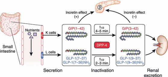
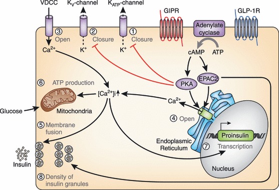
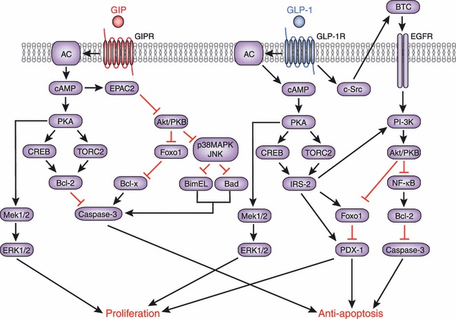
Gastric inhibitory polypeptide (GIP) and glucagon-like peptide-1 (GLP-1) are the two primary incretin hormones secreted from the intestine on ingestion of glucose or nutrients to stimulate insulin secretion from pancreatic β cells. GIP and GLP-1 exert their effects by binding to their specific receptors, the GIP receptor (GIPR) and the GLP-1 receptor (GLP-1R), which belong to the G-protein coupled receptor family. Receptor binding activates and increases the level of intracellular cyclic adenosine monophosphate in pancreatic β cells, thereby stimulating insulin secretion glucose-dependently. In addition to their insulinotropic effects, GIP and GLP-1 play critical roles in various biological processes in different tissues and organs that express GIPR and GLP-1R, including the pancreas, fat, bone and the brain. Within the pancreas, GIP and GLP-1 together promote β cell proliferation and inhibit apoptosis, thereby expanding pancreatic β cell mass, while GIP enhances postprandial glucagon response and GLP-1 suppresses it. In adipose tissues, GIP but not GLP-1 facilitates fat deposition. In bone, GIP promotes bone formation while GLP-1 inhibits bone absorption. In the brain, both GIP and GLP-1 are thought to be involved in memory formation as well as the control of appetite. In addition to these differences, secretion of GIP and GLP-1 and their insulinotropic effects on β cells have been shown to differ in patients with type 2 diabetes compared to healthy subjects. We summarize here the similarities and differences of these two incretin hormones in secretion and metabolism, their insulinotropic action on pancreatic β cells, and their non-insulinotropic effects, and discuss their potential in treatment of type 2 diabetes. (J Diabetes Invest, doi: 10.1111/j.2040-1124.2010.00022.x, 2010).

Keywords: GIP; GLP‐1; Incretin.


Figures

Figure 1
Figure 1
The glucose‐dependent insulinotropic polypepide (GIP) gene is localized on human chromosome 17q21.3–q22 and comprises 6 exons. Proteolytic processing of preproGIP generates GIP that is secreted from K cells. The proglucagon gene is localized on human chromosome 2q36–q37 and comprises 6 exons. In the intestine, proteolytic processing of proglucagon generates glucagon‐like peptide (GLP)‐1 and GLP‐2, whereas glucagon is produced in the pancreas.
Figure 2
Figure 2
Pancreatic and exopancreatic function of glucose‐dependent insulinotropic polypepide (GIP) and glucagon‐like peptide (GLP)‐1. GIP acts directly on the endocrine pancreas, bone, fat, gastrointestinal (GI) tract and brain. GLP‐1 acts directly on the endocrine pancreas, gastrointestinal tract, heart and brain.
Figure 3
Figure 3
Secretion and metabolism of glucose‐dependent insulinotropic polypepide (GIP) and glucagon‐like peptide (GLP)‐1. GIP is secreted from K cells of the upper intestine; GLP‐1 is secreted from L cells of the lower intestine. Released GIP and GLP‐1 rapidly undergoes proteolytic processing by dipeptidyl peptidase‐4 (DPP‐4), and is thereby inactivated and excreted from the kidney. The intact incretins, GIP(1–42), GLP‐1(7–37), and GLP‐1(7–36)amide, have insulinotropic effects on pancreatic β cells, whereas the DPP‐4‐processed incretins, GIP(3–42), GLP‐1(9–37), and GLP‐1(9–36)amide, have lost their insulinotropic effects.
Figure 4
Figure 4
Racial differences in secretion and metabolism of (a) glucose‐dependent insulinotropic polypepide (GIP) and (b) glucagon‐like peptide (GLP)‐1. GIP secretion in healthy Japanese subjects after ingestion of glucose or mixed meals were higher than those of healthy Caucasian subjects, whereas levels of intact GIP were similar37,40–42. This suggests processing of GIP by dipeptidyl peptidase‐4 (DPP‐4) in Japanese subjects might be enhanced. Considerably low levels of intact GLP‐1 after ingestion of glucose or mixed meals are also consistent with enhanced DPP‐4 activities in Japanese subjects. To compare early phase incretin secretion and their processing by DPP‐4 between non‐obese (BMI <25) healthy Japanese and Caucasian subjects, area under the curves during 0–30 min after ingestion of mixed meals containing similar calories are plotted for total and intact forms of (a) GIP and (b) GLP‐1 from Yabe et al.42 Note that total and intact GIP and total GLP‐1 levels were measured in the same immunoassays, whereas assays for intact GLP‐1 were similar except for the presence or absence of the ethanol extraction.
Figure 5
Figure 5
Molecular mechanisms underlying the insulinotropic effects of glucose‐dependent insulinotropic polypepide (GIP) and glucagon‐like peptide (GLP)‐1. Binding of GIP and GLP‐1 to their specific receptors, the GIP receptor (GIPR) and the GLP‐1 receptor (GLP‐1R) leads to activation of adenylate cyclase and subsequent elevation of intracellular cyclic adenosine monophosphate (cAMP) levels. Increased cAMP then activates protein kinase A (PKA) and exchange protein activated by cAMP2 (EPAC2)/cAMP‐guanine nucleotide exchange factor (GEF)II. Activation of PKA promotes closure of KATP channels and facilitates membrane depolarization. PKA also leads to inhibition of the delayed rectifying K+ (Kv) channel, a negative regulator of insulin secretion in pancreatic β cells, resulting in prolongation of action potentials. Depolarization opens the voltage‐gated Ca2+ channels (VDCC), allowing an increase of intracellular Ca2+ concentrations that mobilizes Ca2+ from intracellular stores through PKA‐ and EPAC2‐dependent mechanisms. The increased Ca2+ concentrations eventually trigger fusion of insulin‐containing granules with the plasma membrane and insulin secretion from the β cells. Increased Ca2+ levels also promote transcription of the proinsulin gene, thereby increasing the insulin content of the β cell. Activation of EPAC2 has been shown to increase the density of insulin‐containing granules near the plasma membrane to potentiate insulin secretion from the β cell. ATP, adenosine triphosphate.
Figure 6
Figure 6
Molecular mechanisms underlying the anti‐apoptotic and proliferative effects of glucose‐dependent insulinotropic polypepide (GIP) and glucagon‐like peptide (GLP)‐1. Signaling cascades linking the GIP receptor (GIPR) and the GLP‐1 receptor (GLP‐1R) with anti‐apoptotic and proliferative effects share similarities and differences as shown. Involvement of epidermal growth factor (EGFR) and phosphoinositide 3‐kinase (PI‐3K) has been shown to be a critical difference between the GIPR‐ and GLP‐1R‐signaling pathways. AC, adenylate cyclase; Akt, v‐akt murine thymoma viral oncogene homolog; Bad, Bcl‐2 antagonist of cell death; Bcl, B‐cell CLL/lymphoma; BimEL, Bcl‐2 interacting mediator of cell death EL; BTC, betacellulin; cAMP, cyclic adenosine monophosphate; CREB, cAMP response element‐binding; c‐Src, proto‐oncogene tyrosine‐protein kinase Src; EPAC2, exchange protein directly activated by cAMP2; ERK, extracellular signal‐regulated kinase; Foxo1, forkhead box protein O1; IRS‐2, insulin receptor substrate 2; JNK, c‐Jun N‐terminal kinase; MAPK, mitogen‐activated protein kinase; Mek, mitogen‐activated protein kinase kinase; NFκB, nuclear factor kappa‐light‐chain‐enhancer of activated B cells; PDX‐1, pancreas/duodenum homeobox protein 1; PKA, protein kinase A; PKB, protein kinase B; TORC2, transducer of regulated CREB activity 2.

Similar articles

Cited by

References

    1. Bayliss WM, Starling EH. The mechanism of pancreatic secretion. J Physiol 1902; 28: 325–353 - PMC - PubMed
    1. Moore B. On the treatment of diabetus mellitus by acid extract of duodenal mucous membrane. Biochem J 1906; 1: 28–38 - PMC - PubMed
    1. Zunz E, La Barre J. Contributiona a l’etude des variations physiologiques de la secretion interne du pancreas: Relations entre les secretions externe et interne du pancreas. Arch Int Physiol Biochim 1929; 31: 20–44
    1. Elrick H, Stimmler L, Hlad CJ Jr, et al. Plasma insulin response to oral and intravenous glucose administration. J Clin Endocrinol Metab 1964; 24: 1076–1082 - PubMed
    1. McIntyre N, Holdsworth CD, Turner DS. New interpretation of oral glucose tolerance. Lancet 1964; 2: 20–21 - PubMed

LinkOut - more resources