

Role and mechanism of pancreatic β-cell death in diabetes: The emerging role of autophagy
- PMID: 24843437
- PMCID: PMC4014885
- DOI: 10.1111/j.2040-1124.2010.00054.x
Role and mechanism of pancreatic β-cell death in diabetes: The emerging role of autophagy
Abstract
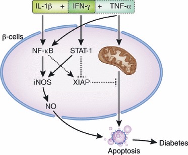
Pancreatic β-cell failure resulting from decreased β-cell mass or dysfunction is the ultimate step towards most types of diabetes. Even if insulin resistance exists, diabetes does not develop unless pancreatic β-cell function or its adaptation is compromised. Classically, two types of cell death (apoptosis and necrosis) have been studied in the diabetes field. Recently, a third type of cell death (autophagy, sometimes called type 2 programmed cell death in comparison with apoptosis, type 1 programmed cell death) and its pathophysiological role have been recognized and are being investigated. In the present review, we will discuss the role of various types of cell death in the development of type 1 and type 2 diabetes. Specifically, we will briefly cover recent progress regarding the role of autophagy in diabetes, which is becoming a hot topic in diabetes and metabolism. (J Diabetes Invest, doi: 10.1111/j.2040-1124.2010.0054.x, 2010).
Keywords: Autophagy; Diabetes; β‐cell death.
Figures
Similar articles
-
Role of pancreatic β-cell death and cell death-associated inflammation in diabetes.Curr Mol Med. 2012 Dec;12(10):1297-310. doi: 10.2174/156652412803833553. Curr Mol Med. 2012. PMID: 22834831 Review.
-
Role of pancreatic β-cell death and inflammation in diabetes.Diabetes Obes Metab. 2013 Sep;15 Suppl 3:141-51. doi: 10.1111/dom.12153. Diabetes Obes Metab. 2013. PMID: 24003931 Review.
-
Autophagy and the pancreatic beta-cell in human type 2 diabetes.Autophagy. 2009 Oct;5(7):1055-6. doi: 10.4161/auto.5.7.9511. Epub 2009 Oct 14. Autophagy. 2009. PMID: 19657235
-
Mitochondrial metabolism and diabetes.J Diabetes Investig. 2010 Oct 19;1(5):161-9. doi: 10.1111/j.2040-1124.2010.00047.x. J Diabetes Investig. 2010. PMID: 24843427 Free PMC article. Review.
-
Apoptosis, autophagy & endoplasmic reticulum stress in diabetes mellitus.Indian J Med Res. 2016 Oct;144(4):515-524. doi: 10.4103/0971-5916.200887. Indian J Med Res. 2016. PMID: 28256459 Free PMC article. Review.
Cited by
-
Defining the ferroptotic phenotype of beta cells in type 1 diabetes and its inhibition as a potential antidiabetic strategy.Front Endocrinol (Lausanne). 2023 Aug 3;14:1227498. doi: 10.3389/fendo.2023.1227498. eCollection 2023. Front Endocrinol (Lausanne). 2023. PMID: 37600723 Free PMC article.
-
Dysfunctions, molecular mechanisms, and therapeutic strategies of pancreatic β-cells in diabetes.Apoptosis. 2023 Aug;28(7-8):958-976. doi: 10.1007/s10495-023-01854-0. Epub 2023 Jun 5. Apoptosis. 2023. PMID: 37273039 Review.
-
Characterizing poorly controlled type 2 diabetes using 1H-NMR metabolomics.Metabolomics. 2024 May 11;20(3):54. doi: 10.1007/s11306-024-02127-w. Metabolomics. 2024. PMID: 38734832 Free PMC article.
-
Targeting ferroptosis: opportunities and challenges of mesenchymal stem cell therapy for type 1 diabetes mellitus.Stem Cell Res Ther. 2025 Feb 4;16(1):47. doi: 10.1186/s13287-025-04188-7. Stem Cell Res Ther. 2025. PMID: 39901210 Free PMC article. Review.
-
A pre-clinical study to investigate the anti-diabetic potential of p-propoxybenzoic acid as a multi-target inhibitor in streptozotocin-nicotinamide induced type-2 diabetic rats.J Diabetes Metab Disord. 2022 Dec 31;22(1):571-580. doi: 10.1007/s40200-022-01177-y. eCollection 2023 Jun. J Diabetes Metab Disord. 2022. PMID: 37255789 Free PMC article.
References
-
- Masini M, Bugliani M, Lupi R, et al. Autophagy in human type 2 diabetes pancreatic beta cells. Diabetologia 2009; 52: 1083–1086 - PubMed
-
- Kim YH, Kim S, Kim KA, et al. Apoptosis of pancreatic beta‐cells detected in accelerated diabetes of NOD mice: no role of Fas‐Fas ligand interaction in autoimmune diabetes. Eur J Immunol 1999; 29: 455–465 - PubMed
-
- O’Brien BA, Huang Y, Geng X, et al. Phagocytosis of apoptotic cells by macrophages from NOD mice is reduced. Diabetes 2002; 51: 2481–2488 - PubMed
-
- Chervonsky AV, Wang Y, Wong FS, et al. The role of Fas in autoimmune diabetes. Cell 1997; 89: 17–24 - PubMed
-
- Kim S, Kim KA, Hwang DY, et al. Inhibition of autoimmune diabetes by Fas ligand: the paradox is solved. J Immunol 2000; 164: 2931–2936 - PubMed
Publication types
LinkOut - more resources
Full Text Sources
